Sunday 30 October 2016

Senescence

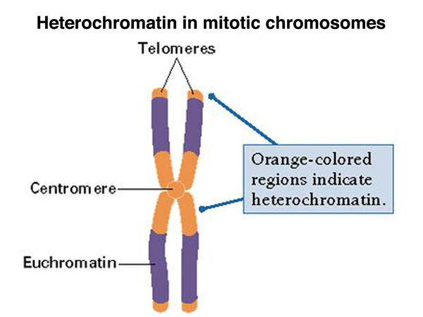
The gradual deteriotion of cell division and growth that lead to death is termed as senescence. Telomeres ends serve to protect the coding DNA of the genome. When a telomeres shorten to critical lengths, the cell senescence and die off. It may affect the whole organism or some parts , cells and tissues. In case of leaf it may be seasonal leaf senescence or sequential leaf senescence.


Role of hormones -
  • Cytokinin is a plant growth regulator which delays leaf senescence by delaying the degradation of chloroplasts . In the leaf it starts from margin and move towards the interior.
  • ABA(Abscissic acid ) induces senescence resulting in chlorosis and necrosis.
Types of senescence-

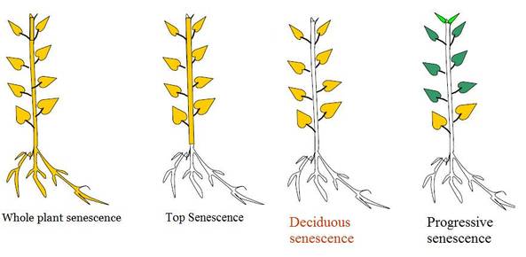
1.Whole plant senescence - Found in  monocarpic plants .Whose are flower only once in their life cycle .(Annuals and bienneals) In this the senescence process begins with reproductive phase and the whole plant dies after seed formation .(Bamboos)

2.Shoot senescence- The upper part of shoot only undergoes senescence. The underground shoot remains as it is . It is seen in perennial plants . Examples zinger ,Musa paradisica 

3.Organ senescence- Occur only in the lateral organs like leaves and fruits . It is of 2 types -

Simultaneous senescence - Seen in deciduous plants where all leaves senesce at a particular season . It is also called as seasonal or deciduous senescence. It is controlled by environmental factors. Example is maple .

Sequential senescence (Progressive) -(Example coleus plant)   As the name indicates the senescence occurs in a sequential manner depending upon the age of leaf. The progressive older and lower leaves senesce while the new ones are added to the shoot .


Image result for plant senescence

                        Image source credit -http://biology4isc.weebly.com/7-plant-hormones-and-photomorphogenesis.html


Thank you for visiting my blog.Please feel free to share your comment on this article ,Please subscribe and share the articles to get more such articles.


Friday 28 October 2016

Stem cells

The cells that  have the capability to give rise to any kind of other cells. It have  totipotent nature that means as in the tissue culture a single explant is able to give rise a new plant the stem cells  can differentiated into other cells and the classification includes -
  • Totipotent - the ability to differentiate into all possible cell types.
  • Pluripotent - the ability to differentiate into almost all cell types. (include embryonic stem cells and cells that are derived from the mesoderm, endoderm, and ectoderm germ layers that are formed in the beginning stages of embryonic stem cell differentiation.)
  • Multipotent - the ability to differentiate into a closely related family of cells.( include hematopoietic (adult) stem cells that can become red and white blood cells or platelets.)
  • Oligopotent - the ability to differentiate into a few cells. Examples include (adult) lymphoid or myeloid stem cells.
  • Unipotent - the ability to only produce cells of their own type, but have the property of self-renewal required to be labeled a stem cell. Examples include (adult) muscle stem cells.
Stem cells can be preserved from the birth time of a baby. Human stem cells are currently being used to test new drugs. New medications are tested for safety on differentiated cells generated from human pluripotent cell lines. 

It can be adult stem cell or embryonic stem cell.

Embryonic stem cells can become all cell types of the body because they are pluripotent. Adult stem cells are thought to be limited to differentiating into different cell types of their tissue of origin.
Embryonic stem cells can be grown relatively easily in culture. Adult stem cells are rare in mature tissues, so isolating these cells from an adult tissue is challenging, and methods to expand their numbers in cell culture have not yet been worked out. This is an important distinction, as large numbers of cells are needed for stem cell replacement therapies. 


Thank you for visiting my blog.Please share your comments. Please subscribe and share the articles . Follow me on Google + to get notified with new articles.

Friday 21 October 2016

Bombay blood group

U might have thinking that  why the name of the blood group is Bombay blood group ? Is this only found in people who are living in Bombay ?

It is called as Bombay blood group as this blood group is first identified in Bombay by Dr.Y.M.Bhende in 1952 . It is also called as  HH group . The peculiarity is that this type of blood group do not express the H antigen. As a result they cannot form A antigens or B antigens on their red blood cells. Thus they can donate blood to anybody but can receive blood only from Bombay blood group people. Any person with this blood group who needs an urgent blood transfusion will probably be unable to get it, as no blood bank would have any in stock.

Some movies are also made by taking this concept of Bombay blood group . It is a very rare blood type . Sometimes people get confused between o blood group and Bombay blood group as both can donate their blood to all other blood types and it also lack both antigens a and b . The difference is that in case of O blood group Fucose is present and H antigen is present .

It can be Rh positive or Rh negetive .Try to understand the concept when we say someone has A blood group it means it contain Ag of blood group A and Ab of type B in the blood . People with AB blood group has both Ag a and Ag b but no antibodies. Blood group O has both A and B Antibodies but no antigens .It is not well known that all have antigen H .So, it is found in people with O +ve blood group where antigen H is missing ( Bombay blood group).


Image result for bombay blood group

Image source credit -https://rhesusnegativebloodgroup.files.wordpress.com/2011/10/toi_2010_8_21_5.jpg
MCQS - 

1.About Bombay blood group, true is
A. Lack of A, B and H antigen's on RBC
B. Lack of only A and B antigens
C. Only A
D. Both A and B
Ans.  A
Thank you for visiting my blog.Please feel free to share your comment on this article ,Please subscribe and share the articles to get more such articles.

Monday 3 October 2016

Heterochromatin And Euchromatin

Heterochormatin - It is a region of chromosome whose properties are as follows-
  • It is highly condensed.
  • It is found in centromere and telomere region .
  • It contains long stretches of repetitive sequences called satellite DNA.
  • Replication occours in late s- phase .
  • Meiotic recombination is absent .
  • Heterochomatin Dna is not very active.
  • Transcriptionally silent and silences adjecent genes .
  • Types of heterochromatin -
1.Constitutive Heterochromatin - It remains always as heterochromatin untill no change due to mutation hence also called as obligate heterochromatin. It is usually repetitive and forms structural functions such as centromere or telomers.
e.gCentromeric heterochromatin,telomeric heterochromatin, Retrotransposons

2.Facultative Heterochromatin -It may convert to euchromatin depending upon the requirement .It is the result of genes that are silenced through a mechanisms such as Histone methylation or sirna by RNAi.
e.g Barr body in Mammalian female and female Drosophila 


Image result for heterochromatin
                                    Image source credit -http://www.discoveryandinnovation.com/BIOL202/notes/lecture18.html

Euchromatin -
  • It is less condensed ,loosely packed regions of chromatin .
  • It is lightly stained.
  • Replication occours in throughout  s- phase .
  • Meiotic recombination is present.
  • Euchomatin Dna is very active.
  • Transcriptionally  not silent, very active .
MCQs.
1.In eukaryotes, transcription is generally associated with 
A) euchromatin only. 
B) heterochromatin only. 
C) very tightly packed DNA only. 
D) both euchromatin and histone acetylation

Ans. d

2.Some of the heterochromatin region can convert into euchromatic regions ,in different cell cycle stages. They are called -
a.Constitutive heterochromatin
b.Facultative heterochromatin
c.Euchromatin 
d.None of the above 

Ans. b
Thank you for visiting my blog.Please feel free to share your comment on this article ,Please subscribe and share the articles to get more such articles.

Collagen

It is a type of structural protein which is most abundant protein in the human body . It is found throughout the body . It is the main fibrous component of skin , tendon,bone and cartilage. Normal collagen denatures at 39 c and on boiling they yield gelatin .Vertebrates have 46 genetically distinct polypeptide chains comprising 28 distinct collagen types . it is a secreted protein and therefore occurs outside cells. They are hydrolyzed by pepsin Enzyme. 

Structure -triple helix structure (3.3  aminoacids residues per turn )
  • Collagens are made up of 3 polypeptide alpha chains which are left handed and coiled around each other to form a right alpha helix confirmation . 
  • Each such helix is around 1.4 nm in diameter and 300 nm in lengthThe rise of the collagen helix (superhelix) is 2.9 Å (0.29 nm) per residue. 
  • Glycine occupies every third position in the repeating amino acid sequence. (Gly-X-Y). X is often proline. Proline makes up about 17% of collagen.
  • Collagen contains two uncommon derivative amino acids . These amino acids are found at specific locations relative to glycine and are modified post-translationally by different enzymes, both of which require vitamin-C as a co-factor-
  • 1.Hydroxyproline derived from proline .     2. Hydroxylysine derived from lysine.
  • Proline and hydroxyproline residues permit sharp twisting of collagen helix.Only glycine residues can be accomodated at tight junctions between chains.

Formation of fibril -

1.After the formation of alpha chain the sequence facilitate binding of ribosomes to the rough ER and direct the alpha chain into the lumen of the RER.

2.This sequence is cleaved and precurser of collagen Pro- alpha chain is formed.
3.Proline and lysine are hydrolysed and modified by glycosylation with glucose or galactose residues.

4.After hydroxylation and glycosylation pro alpha chains are converted to pro -collagen .

5.Pro collagens are translocated to Golgi - apparatus .

6.In the golgi apparatus they are packaged in secretory vesicles or transport vesicles (Exocytosis).

7.Vesicles fuse with the membrane and release the pro- collagen into extracellular space.

8. Procollagen molecules are cleaved by N and C pro collagen peptides.

9.Triple helical structure is released as Tropocollagen .

10.Tropocollagen spontaneously associate with each other and form collagen fibrils .
Thank you for visiting my blog.Please feel free to share your comment on this article ,Please subscribe and share the articles to get more such articles.

Friday 30 September 2016

Genomic imprinting

Genomic imprinting is an epigenetic (study of changes in organisms caused by modification of gene expression rather than alternation of the genetic code itself )phenomenon which in most cases is belived to occur in gametogenesis.

 It occurs when both maternal and paternal alleles are present ,but one allele will be expressed while the other remains inactive.This process is necessary for development and may somehow regulate growth in the embryo .

Evidence - It came from experiments with androgenotes ( embryos with two paternal genomes ) and gynogenotes ( embryos with two maternal genomes ) , which were produced by nucler transplantation .These zygotes are formed , but neither type was able to undergoes further development.

Example -
1. Gene Igf2 is an imprinted gene being expressed only from the paternal chromosome .
2. H19 gene is an imprinted gene expressed only from the maternal chromosome .  


Image result for genomic imprinting
                                                       Image source credit-http://curekatelyn.com/how-close-are-we-to-a-cure/


It is also called as uniparental disomy as only one allele is expressed either from father or from mother .

Genomic imprinting is rare in mammals because most of the genes are not imprinted. From a nuclear transplantation experiment in mouse during  1980 s it was confirmed that foe normal mammalian development both the maternal and paternal genomes are required.

In case of flowering plants some genes are found to be expressed from maternal genomes while some others are expressed exclusively from the alone paternal copy .( In case of Sweetbay -Magnolia virginiana )

In case of insects like honeybees and  ants can reproduce either through fertilized eggs or unfertilized eggs. This sex determining system develops males from unfertilized eggs and females develop from fertilized eggs. The queen honey bee decides whether to fertilize an egg or to leave it unfertilized .

Improper imprinting in humans leads to some developmental abnormalities-
1.Prader-Willi-syndrome (when fathers copy is missing or there are 2 maternal parents.)
2.Angelman syndrome( when mothers copy is missing and there are 2 paternal parents .)


Thank you for visiting my blog.Please feel free to share your comment on this article ,Please subscribe and share the articles to get more such articles.

Thursday 29 September 2016

Hypervolume niche

Hypervolume Niche- The limits, for all important environmental conditions, within which individuals of a species can survive, grow and reproduce is termed as niche . If each condition, resource or other species is seen as a dimension, the niche is a n-dimensional hypervolume. 

Types of hypervolume niches-

 a. Fundamental niche 
 b. Realized. niche

 Fundamental niche is the the actual entire niche that an oraganism/ species occupy ,when species is not competing with others for its resources. But individual or a species normally remains in competition (either interspecific or intraspecific or both) and under such conditions, only a part of the niche is realized by the species. This smaller hypervolume occupied by the species is called realized niche.

Thus,each species has both fundamental niche within a community to which it is adapted in the evolutionary process, but because of competition it occupies a smaller niche, namely realized niche.
 In  competing zone due to competition, the reproductive success of each species and hence chances of survival reduced.

Individuals, who remain outside the overlapping competitive zone, are likely to have a greater survival rate and reproductive success.Natural selection favors individuals lying in the non-competing zone.

The fundamental niche is larger then the realized niche. The realized niche can be called a subset of the fundamental niche. 


Image result for fundamental niche

                                 Imge source credit -https://quizlet.com/75558972/bio-lecture-17-flash-cards/

When all of the boundaries of tolerance of diverse environmental influences are assembled into a single, multivariate factor. This is known as  the multidimensional zone (also known as a hypervolume) of environmental tolerance, in which an individual can potentially survive or in which a species can maintain viable populations. We can quantify in this the niche breadth and niche overlap .


Thank you for visiting my blog.Please feel free to share your comment on this article ,Please subscribe and share the articles to get more such articles.

Wednesday 28 September 2016

Transgenic plants

Transgenic plants are genetically modified plants by introduction or expression of a foreign gene. It is also called as genetically engineered plants. It use the recombinant Dna technology  so we can say it is a result of modern biotechnology .

Production of transgenic plants -

1. Isolation - isolate and clone the gene of intrest. The gene is called transgene.
2.Addition of segments- Add the Dna segments for the initiation of the gene expresiion.
3.Selectable markersSelectable markers are those which allow the selection of transformed cells, or tissue explants, by their ability to grow in the presence of an antibiotic or a herbicide. The most frequently used selectable markers are kanamycin and hygromycin. In addition to selecting for transformants, such markers can be used to follow the inheritance of a foreign gene in a segregating population of plants.
4.Transformation -Transfer the gene construct into plant cell .
5.Selection of transfered cells or tissues .
6.Regenerate a new whole plant .

Advantage -
1. It improves the size of nutrients.
2.Reduce the percentage of waste gives a longer shelf life .
3.Plant cells are totipotent in nature that means a whole plant can generate  from an explant .
3. Increase the yield of crops
4.Improves the resistant power against disease,pests and herbicides.
5.Decrease pollution -Some genetically modified plants are made to prevent heavy metal pollution from contaminated soil.
6.Tolerance to drought and salinity .

Examples-

1.Bt corn varieties (for pest resistance ) 1995 for example, contain a gene from a bacterium (Bacillus thuringiensis) found in the soil that causes the transgenic corn to produce an insecticidal protein. The gene confers resistance to ball worms.

2.Slow ripening tomato (Flavr savr )1994 It is the first commersialized GM crop .
  • It improves the texture.
  • It delays the ripening process.
  • It helps in facilitation of transportation .
3.Golden rice ( Improves nutritional quality )

4.GM maize (drought tolerance)- Transgenic maize expresses a Rna chaperone gene in tolerant to salt and water stress . 
1.
Q. The transgenic animals are those which have
(a) foreign DNA in some of its cells
(b) foreign DNA in all its cells
(c) foreign RNA in all its cells
(d) DNA and RNA both in the cells
Ans.b
Thank you for visiting my blog.Please feel free to share your comment on this article ,Please subscribe and share the articles to get more such articles.

Monday 26 September 2016

Gastrulation

It is the process of cell and tissue movements whereby the cells of the blastula are rearranged in different manner.

  • The blastula consists of numerous cells , the positions are established during cleavage.
  • The three germ layers such as ectoderm(outside), endoderm and mesoderm are first produced in the process of gastrulation .
  • Skin and nervous system system are spread over its outside surface.
  • The movements of gastrulation involve the entire embryo and cell migrations in one part of the gastrulating embryo which  coordinate with other movements occuring simultaneously.
Gastrulation usually involves some combination of the following types of movements -

1. Invagination - (sea urchin endoderm)The infolding of a cells ,much like the indenting of a soft rubber bait when it is pocked .(inward buckling of an epithelium.)

Image result for invagination of embryo
                                Image source credit-https://www.studyblue.com/notes/note/n/developmental-bio-exam-1/deck/5569140

2.Involution - (Amphibian mesoderm) The inward movement of an expanding outer layer so that it spreads over the internal surface of the remaining external cells. It requires migration .(movement of individual cells over other cells ).

Image result for involution of embryo
                                     Image source credit-http://biology.kenyon.edu/courses/biol114/Chap14/Chapter_14.html


3.Ingression - (Sea urchin mesoderm and Drosophila neuroblasts) Migration of individual cells from the surface layer into the cativy or interior of the embryo .
Image result for ingression  of embryo
                             Image source credit -http://www.biology.arizona.edu/developmental_bio/problem_sets/developmental_mechanisms/03t.html

4.Delamination -( Mammalian and bird hypoblast formation ) Spilliting of 1 cell sheet into 2 or more parallel sheets. 

5.Epiboly- (Ectoderm formation in amphibians, sea urchins and tunicates )Spread of an outside cell layer(epidermal sheets) to envelop a yolk mass or deeper layer.or expansion of one cell sheet over other cells.

Image result for delamination of embryo
Types of cell movements during gastrulation 
                                  Image source credit -http://courses.biology.utah.edu/bastiani/3230/DB%20Lecture/Lectures/a7Gast.html


Thank you for visiting my blog.Please feel free to share your comment on this article ,Please subscribe and share the articles to get more such articles.

TWO PIGMENT SYSTEMS

Two Pigment Systems: 

In result of red drop and Emerson effect it was concluded that at least two pigment systems are involved in photosynthesis. These two pigment systems are -

1. PS1 (piment system 1)

2.PS2(pigment system 2)

Presence of two such systems has been supported by studies based on chloroplast fractionation process which showed two types of particles within the chloroplast membrane. Acc . to Salisbury and Ross in 1986 smaller and lighter particles of PS I(located in non-appressed regions (stroma lamella) of  thylakoids) and larger and heavier particles of PS II(in appressed regions (grana ) of thylakoids ).

PS I complex -

  • Consists of 200 chlorophylls, 50 carotenoids and  a molecule of P 700.
  • Cytochrome f, one plastocyanin, two cyt-b, FRS (ferredoxin reducing substance), one or two membrane bound ferredoxin molecules etc.are also present.
  • Primary electron acceptor is Fd .
  • The carotenoids collect light energy for both the systems. Reduced carotenoids such as carotenes are found in PS-I. 
  • It is rich in chl-a, iron and copper.  PS I controls the process of producing a strong reductant to reduce NADP into NADPH + H+.
  • It involves in cyclic as well as non cyclic phosphorylation .
  • It is not associated with photolysis of water .
  • Main function is ATP sunthesis and production of NADPH.


Z-scheme of elctron transport chain 
Image source credit -http://www.bio.miami.edu/dana/226/226F08_10print.html

PSII complex -
  • Consists of 200 chlorophylls, 50 carotenoids, a molecule of P 680, and a primary electronacceptor Q as plastoquinone, four plastoquinone equivalents four Mn++molecules bound to one or more proteins, two cyt-b6 559, one cyt-b6 and chloride. 
  •  PS II is concerned with generation of strong oxidant and weak reductant coupled with the release of oxygen.
  • More oxidised forms as xanthophylls, violaxanthin and neoxanthin are found in PS-II. 
  • It involves in both non-cyclic phosphorylation .
  • It is associated with photolysis of water.
Image result for photolysis of water
(water molecules lyse using light energy, produces H+ ions (protons) e- (electrons) and oxygen. )
                                                 Image source credit -http://www.memrise.com/mem/2084591/photolysis-of-water/
  • Main function is ATP synthesis and hydrolysis of water.
Thank you for visiting my blog.Please feel free to share your comment on this article ,Please subscribe and share the articles to get more such articles.

Sunday 25 September 2016

Aquaporins

  Image result for aquaporins diagram

Image source credit- https://www.studyblue.com/notes/note/n/bio-111-study-guide-2012-13-wan/deck/9724270


Aquaporins are membrane proteins of a cell which helps in regulating the flow of water. It is a 28Kda protein . This is a kind of water channels which facilitate the movement of water by incresing the permiability of water towards membrane . It is found in all kingdom of life like plants, animals and micro-organisms.

The structure of Aquaporin is made up of 6 membrane helices.

Functions -
1. In cell migration .
2.Skin hydration . (AQP3 &AQP7)
3.Cell proliferation particularly AQP3.
4.AQP7 found to be involved in fat metabolism .
5.Conduct water molecules in and out of the cell and preventing the flow of ions and other molecules.
6. AQPs have also been proposed to transport other small molecules and gases, including carbon dioxide, ammonia, nitric oxide and hydrogen peroxide .
7.The AQP4 water channel is expressed in astrocytes throughout the central
nervous system.

The strucuture of aquaporins have been defined by mutagenesis, spectroscopy and freeze fracture electron microscopy method.
                                          CSIR QUESTIONS                Q.Which of the following statements describe the characteristics of  an aquaporin-

A)they are membrane proteins that are soluble in water. 
B)they are protein molecules with channels through which water molecules can diffuse.
C)their numbers in a membrane can change due to differing conditions or hormonal stimulation.
D)B& C

Ans. (D) They are actually hydrophobic proteins and therefore not soluble in water. Aquaporins are especially abundant in kidney and intestinal epithelial cells where water transport occurs extensively across tubular and epithelial cells. Dehydration condition can stimulate the kidney tubules to increase the number of water channels under stimulation by the posterior pituitary hormone ADH.

Q2.Aquaporins are a class of proteins that are relatively abundant in plant membranes .Following are certain statements -
a.Aquaporins form water channels in membrane.
b.some aquaporins also transport uncharge molecules such as ammonia.
c.Activity of aquaporins is not regulated by phosphorylation.
d.Activity is regulated bu calcium concentration and reactive oxygen species .
Which of the combinations are correct -
a.A,B& D                    C.A,C&D
b.B,C &D                          D.A,B& C

Ans. a (A,B &D ) the aquaglyceroporins transport uncharged molecules such as ammonia,urea,glycerol and CO2.

NOTE- Aquaporin proteins are composed of a bundle of six transmembrane α-helices. They are embedded in the cell membrane. The amino and carboxyl ends face the inside of the cell. The amino and carboxyl halves resemble each other, apparently repeating a pattern of nucleotides. Some researchers believe that this was created by the doubling of a formerly half-sized gene. Between the helices are five regions (A – E) that loop into or out of the cell membrane, two of them hydrophobic (B, E), with an asparagine–proline–alanine ("NPA motif") pattern. They create a distinctive hourglass shape, making the water channel narrow in the middle and wider at each end.


Thank you for visiting my blog.Please feel free to share your comment on this article ,Please subscribe and share the articles to get more such articles.

Thursday 22 September 2016

Fats and Oils

Fats, oils and waxes are the general terms describing a structurally diverse bio-macromolecule called lipids. Lipids are esters of glycerol and fatty acids. Lipids with saturated fatty acids (saturated fat) tend to be solid at room temperature (25oC) and hence we generally call it as ‘fat’. On the other hand, lipids with unsaturated fatty acids (unsaturated fat) tend to be liquid at room temperature and thus they are called ‘oils’. Lipids are present in all living organisms including Archaebacteria. 
Image result for fats and oils structure

                    Imagesourcecredit-http://images.flatworldknowledge.com/ballgob/ballgob-fig17_x009.jpg

Similarities -
1. Both animal and plant fats are triglycerides (one glycerol esterified with three fatty acid residues)
2. Both are food reserve of the cell
3.  Unsaturated and saturated fats occurs in both plants and animals .
Difference -
Animal fats (Fats) Butter fat, Beef fat
  •  Animals fats are relatively rich in saturated fatty acids.
  • Animals fats due to their saturation, tend to stay solid at room temperature, hence commonly known as ‘fats’.
  • Iodine number of animal fats will be relatively less (iodine number denote the degree of un-saturation in fatty acids).
  •  Oxidative rancidity is observed more frequently in animal fats.
  •  Animal fats are stored in liver, beneath the skin etc.
  •  Animals have specialized cells for storing fat reserve called adipocytes.
  • Vanderwall forces between the molecules are stronger .

Plant fats or Vegetable fats (Oil ) Coconut oil, Olive oil, Sunflower oil
  • Plant fats are comparatively rich in unsaturated fatty acids.
  •  Plant fats due to their high un-saturation, tend to stay liquid at room temperature, hence commonly known as ‘oils’.
  •  Iodine number of plant fats will be more when compared to animal fats.
  •  Oxidative rancidity is relatively less in plant fats.
  •  Plant fats are stored in fruits and seeds.
  •  Plants do not have adipocytes for storing fats, fats are stored as granules (oil droplets) in the cell cells of endosperm of seeds or other cells.
  • Vanderwall forces between the molecules are weaker.

Thank you for visiting my blog.Please feel free to share your comment on this article ,Please subscribe and share the articles to get more such articles.

Tuesday 20 September 2016

Archaebacteria

A kind of bacteria which can live in very harsh environment . They do not require oxygen and they can sustain in very hot condition ,extreme salty enviornment and deep sea hydrothermal vents  . They are unicellular prokaryotes .


Image result for archaebacteria classification

                     Image source credit -http://lady-rosales.blogspot.in/2010/08/three-domains-system-three-domain.html


Methanophiles (in sewage plant to help in breakdown of sewage ), halophiles (high conc. of salt ) and thermophiles (extreme hot areas ) are the examples of archaebacteria. These are looks like bacteria in microscope .

It differs from eubacteria in the respect of  rna which is present in ribosomes and arranged differently. They are thought to be oldest living organisms on earth .

Characters -

1. It stain  Gram + ve or -ve .
2. shape - sperical, lobed, spiral, rod shape
3. multiplication is asexual by binary fission or budding or fragmentation .
4. Circular DNA like bacteria and lacks histone protein .
5.GΨC content varies from 21-68%.
6. in T C arm of t- rna - pseudouridine/ 1- methyl pseudo uridine is present .
7. I t is sensitive to anisomycin and insensitive to kanamycin and chloramphenicol.
8. EF-2 reacts with diptheria toxin .
9. It obtain energy from sulphur and ammonia.
10. Methanogens live in deep mud without oxygen .
11.Halophiles can utilize light energy for the direct synthesis of ATP.
12.It is responsible for producing methane in bio gas ferementer.
13.Cell walls made up principally of pseudomurein.( N-acethyltalosaminuronic acid)Lacks D amino acids.
14.Ether linkage is present instead of ester linkage.
15.RNA pol enzyme contains 5-11 different subunits .


MCQ. 
Q.Archaebacteria differ from eubacteria in
1.cell membrance structure
2.mode of nutrition
3.cell shape
4.mode of reproduction

ans. 1


Thank you for visiting my blog.Please share your comments . Subscribe and share the articles and follow me on Google + to get notified about new articles.