Sunday, 30 October 2016

Senescence

The gradual deteriotion of cell division and growth that lead to death is termed as senescence. Telomeres ends serve to protect the coding DNA of the genome. When a telomeres shorten to critical lengths, the cell senescence and die off. It may affect the whole organism or some parts , cells and tissues. In case of leaf it may be seasonal leaf senescence or sequential leaf senescence.


Role of hormones -
  • Cytokinin is a plant growth regulator which delays leaf senescence by delaying the degradation of chloroplasts . In the leaf it starts from margin and move towards the interior.
  • ABA(Abscissic acid ) induces senescence resulting in chlorosis and necrosis.
Types of senescence-

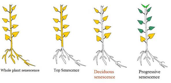
1.Whole plant senescence - Found in  monocarpic plants .Whose are flower only once in their life cycle .(Annuals and bienneals) In this the senescence process begins with reproductive phase and the whole plant dies after seed formation .(Bamboos)

2.Shoot senescence- The upper part of shoot only undergoes senescence. The underground shoot remains as it is . It is seen in perennial plants . Examples zinger ,Musa paradisica 

3.Organ senescence- Occur only in the lateral organs like leaves and fruits . It is of 2 types -

Simultaneous senescence - Seen in deciduous plants where all leaves senesce at a particular season . It is also called as seasonal or deciduous senescence. It is controlled by environmental factors. Example is maple .

Sequential senescence (Progressive) -(Example coleus plant)   As the name indicates the senescence occurs in a sequential manner depending upon the age of leaf. The progressive older and lower leaves senesce while the new ones are added to the shoot .


Image result for plant senescence

                        Image source credit -http://biology4isc.weebly.com/7-plant-hormones-and-photomorphogenesis.html


Thank you for visiting my blog.Please feel free to share your comment on this article ,Please subscribe and share the articles to get more such articles.


Friday, 28 October 2016

Stem cells

The cells that  have the capability to give rise to any kind of other cells. It have  totipotent nature that means as in the tissue culture a single explant is able to give rise a new plant the stem cells  can differentiated into other cells and the classification includes -
  • Totipotent - the ability to differentiate into all possible cell types.
  • Pluripotent - the ability to differentiate into almost all cell types. (include embryonic stem cells and cells that are derived from the mesoderm, endoderm, and ectoderm germ layers that are formed in the beginning stages of embryonic stem cell differentiation.)
  • Multipotent - the ability to differentiate into a closely related family of cells.( include hematopoietic (adult) stem cells that can become red and white blood cells or platelets.)
  • Oligopotent - the ability to differentiate into a few cells. Examples include (adult) lymphoid or myeloid stem cells.
  • Unipotent - the ability to only produce cells of their own type, but have the property of self-renewal required to be labeled a stem cell. Examples include (adult) muscle stem cells.
Stem cells can be preserved from the birth time of a baby. Human stem cells are currently being used to test new drugs. New medications are tested for safety on differentiated cells generated from human pluripotent cell lines. 

It can be adult stem cell or embryonic stem cell.

Embryonic stem cells can become all cell types of the body because they are pluripotent. Adult stem cells are thought to be limited to differentiating into different cell types of their tissue of origin.
Embryonic stem cells can be grown relatively easily in culture. Adult stem cells are rare in mature tissues, so isolating these cells from an adult tissue is challenging, and methods to expand their numbers in cell culture have not yet been worked out. This is an important distinction, as large numbers of cells are needed for stem cell replacement therapies. 


Thank you for visiting my blog.Please share your comments. Please subscribe and share the articles . Follow me on Google + to get notified with new articles.

Friday, 21 October 2016

Bombay blood group

U might have thinking that  why the name of the blood group is Bombay blood group ? Is this only found in people who are living in Bombay ?

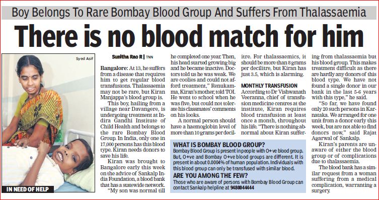
It is called as Bombay blood group as this blood group is first identified in Bombay by Dr.Y.M.Bhende in 1952 . It is also called as  HH group . The peculiarity is that this type of blood group do not express the H antigen. As a result they cannot form A antigens or B antigens on their red blood cells. Thus they can donate blood to anybody but can receive blood only from Bombay blood group people. Any person with this blood group who needs an urgent blood transfusion will probably be unable to get it, as no blood bank would have any in stock.

Some movies are also made by taking this concept of Bombay blood group . It is a very rare blood type . Sometimes people get confused between o blood group and Bombay blood group as both can donate their blood to all other blood types and it also lack both antigens a and b . The difference is that in case of O blood group Fucose is present and H antigen is present .

It can be Rh positive or Rh negetive .Try to understand the concept when we say someone has A blood group it means it contain Ag of blood group A and Ab of type B in the blood . People with AB blood group has both Ag a and Ag b but no antibodies. Blood group O has both A and B Antibodies but no antigens .It is not well known that all have antigen H .So, it is found in people with O +ve blood group where antigen H is missing ( Bombay blood group).


Image result for bombay blood group

Image source credit -https://rhesusnegativebloodgroup.files.wordpress.com/2011/10/toi_2010_8_21_5.jpg
MCQS - 

1.About Bombay blood group, true is
A. Lack of A, B and H antigen's on RBC
B. Lack of only A and B antigens
C. Only A
D. Both A and B
Ans.  A
Thank you for visiting my blog.Please feel free to share your comment on this article ,Please subscribe and share the articles to get more such articles.
Monday, 3 October 2016

Heterochromatin And Euchromatin

Heterochormatin - It is a region of chromosome whose properties are as follows-
  • It is highly condensed.
  • It is found in centromere and telomere region .
  • It contains long stretches of repetitive sequences called satellite DNA.
  • Replication occours in late s- phase .
  • Meiotic recombination is absent .
  • Heterochomatin Dna is not very active.
  • Transcriptionally silent and silences adjecent genes .
  • Types of heterochromatin -
1.Constitutive Heterochromatin - It remains always as heterochromatin untill no change due to mutation hence also called as obligate heterochromatin. It is usually repetitive and forms structural functions such as centromere or telomers.
e.gCentromeric heterochromatin,telomeric heterochromatin, Retrotransposons

2.Facultative Heterochromatin -It may convert to euchromatin depending upon the requirement .It is the result of genes that are silenced through a mechanisms such as Histone methylation or sirna by RNAi.
e.g Barr body in Mammalian female and female Drosophila 


Image result for heterochromatin
                                    Image source credit -http://www.discoveryandinnovation.com/BIOL202/notes/lecture18.html

Euchromatin -
  • It is less condensed ,loosely packed regions of chromatin .
  • It is lightly stained.
  • Replication occours in throughout  s- phase .
  • Meiotic recombination is present.
  • Euchomatin Dna is very active.
  • Transcriptionally  not silent, very active .
MCQs.
1.In eukaryotes, transcription is generally associated with 
A) euchromatin only. 
B) heterochromatin only. 
C) very tightly packed DNA only. 
D) both euchromatin and histone acetylation

Ans. d

2.Some of the heterochromatin region can convert into euchromatic regions ,in different cell cycle stages. They are called -
a.Constitutive heterochromatin
b.Facultative heterochromatin
c.Euchromatin 
d.None of the above 

Ans. b
Thank you for visiting my blog.Please feel free to share your comment on this article ,Please subscribe and share the articles to get more such articles.
Collagen

It is a type of structural protein which is most abundant protein in the human body . It is found throughout the body . It is the main fibrous component of skin , tendon,bone and cartilage. Normal collagen denatures at 39 c and on boiling they yield gelatin .Vertebrates have 46 genetically distinct polypeptide chains comprising 28 distinct collagen types . it is a secreted protein and therefore occurs outside cells. They are hydrolyzed by pepsin Enzyme. 

Structure -triple helix structure (3.3  aminoacids residues per turn )
  • Collagens are made up of 3 polypeptide alpha chains which are left handed and coiled around each other to form a right alpha helix confirmation . 
  • Each such helix is around 1.4 nm in diameter and 300 nm in lengthThe rise of the collagen helix (superhelix) is 2.9 Å (0.29 nm) per residue. 
  • Glycine occupies every third position in the repeating amino acid sequence. (Gly-X-Y). X is often proline. Proline makes up about 17% of collagen.
  • Collagen contains two uncommon derivative amino acids . These amino acids are found at specific locations relative to glycine and are modified post-translationally by different enzymes, both of which require vitamin-C as a co-factor-
  • 1.Hydroxyproline derived from proline .     2. Hydroxylysine derived from lysine.
  • Proline and hydroxyproline residues permit sharp twisting of collagen helix.Only glycine residues can be accomodated at tight junctions between chains.

Formation of fibril -

1.After the formation of alpha chain the sequence facilitate binding of ribosomes to the rough ER and direct the alpha chain into the lumen of the RER.

2.This sequence is cleaved and precurser of collagen Pro- alpha chain is formed.
3.Proline and lysine are hydrolysed and modified by glycosylation with glucose or galactose residues.

4.After hydroxylation and glycosylation pro alpha chains are converted to pro -collagen .

5.Pro collagens are translocated to Golgi - apparatus .

6.In the golgi apparatus they are packaged in secretory vesicles or transport vesicles (Exocytosis).

7.Vesicles fuse with the membrane and release the pro- collagen into extracellular space.

8. Procollagen molecules are cleaved by N and C pro collagen peptides.

9.Triple helical structure is released as Tropocollagen .

10.Tropocollagen spontaneously associate with each other and form collagen fibrils .
Thank you for visiting my blog.Please feel free to share your comment on this article ,Please subscribe and share the articles to get more such articles.