Tuesday, 28 February 2017

INTERFERONS


 Interferons are present in birds, reptiles and fishes as well as higher animals.There are 3 main families of interferons: ab and gGamma interferon is a cytokine which is characteristically a product of Th1-cells and natural killer (NK) cells.

1.Interferons are 
a) anti bacterial proteins 
b) anti-viral proteins
c) bacteriostatic proteins 
d) all of these
Ans.b

2.Antiviral glycoproteins released by living cells in response to viral attack and induce a viral resistant state to neighbouring cells is called as 
a) natural killer cells 
b) complement system 
c) interferons 
d) phagocytes
Ans.c

3. Interferons:
  Are found only in mammalian species.
  Are divided into 5 main families.
  Induce enzyme synthesis in the target cell.
 Are specific for individual viruses.
Correct.  answer -C  Derepression of genes in the target cell result in the synthesis of a protein kinase and an endonuclease.


Thank you for visiting my video tutoria/ blog.Please feel free to share your comment on this
article ,Please subscribe and share the articles to get more such articles.

Monday, 27 February 2017

PAPAIN AND PEPSIN DIGESTION OF IMMUNOLOGY

MCQs-
1.Papain digest IgG into-
A. two Fab fragments and one Fc fragment
B. three Fab fragments and two Fc fragments
C. two Fab fragments and two Fc fragments
D. three Fab fragments and three Fc fragments
Ans.A

2. Papain is found in
A. papaya
B. proteases
C. pepsin
D. pancreatic amylase
Ans. 1

3.Which enzymres are in the composition of gastric juice ?

(A) Ptylin, Renin, Lipase
 (B) Pepsin, Ptylin, Renin
(C) Lipase Pepsin, Renin
(D) Ptylin, Lipase,Pepsin
Ans. C
4.Pepsin has an optimum pH of:
a) 1
b) 1.2
c) 1.4
d) 1.6
Ans. d
Note-Enzyme papain cleaves the immunoglobulin at the N-terminal side of the inter-heavy chain disulfide bond and produces three fragments (two Fab fragments and one Fc fragment).

Enzyme pepsin cleaves the immunoglobulin at the C-terminal region and produces one large fragment (which contains two Fab regions) called F (ab’) 2. The remaining heavy chains on the C-terminal side are digested into multiple small peptides.

4. Cleavage of IgG by papain produces:
  Divalent antigen binding fragments.
  Isolated light chains.
  F(ab')2.
D  Fab.

Ans D  Papain splits the hinge sequences above the inter-heavy chain disulfide bond and produces a monovalent antigen binding fragment consisting of light chain and part of the heavy chain containing the VH and CH1 domains.


Thank you for visiting my video tutorial/blog.Please feel free to share your comment on this
article ,Please subscribe and share the articles to get more such articles.

Friday, 24 February 2017

ANTIGEN ANTIBODY INTERACTION BONDS

MCQs-

1.The bonds involved in antigen-antibody interactions are
a) weak hydrogen bonds and vanderwalls forces
b) strong covalent bonds
c) strong di- sulphide bonds
d) all of these
Ans. a


2.Binding of antigen to antibody:
A.   Is usually unaffected by molecular rigidity. 
B. Is unaffected by the presence or absence of water molecules.
C.  Involves covalent bonding.
D.   Is optimized by spatial complementarity.


Ans. D Spatial complementarity is vital for strong binding of antigen to antibody otherwise non-specific intermolecular forces which depend reciprocally on distance apart, can become really strong.

3.The intermolecular forces which contribute to the interaction between antibody and antigen:
  Are all electrostatic.
  Are all vanderWaals.
  Are all hydrophobic.
 D  Are all hydrogen bonds.
  Rely on a combination of the above.


Ans.E Antibody–antigen interactions depend upon a combination of intermolecular forces, all of which are non-covalent.

Note-
1.The typical length of a hydrogen bond in water is 197 pm. 
2.The atom that looses the electron acquires positive charge and the other atom which gains the electron becomes a negatively charged particle. 
3.Vanderwall forces are weak forces of attraction between molecules. These forces decrease as the molecule gets smaller and increase as the molecule increases. 

Vanderwalls force ~molecular size 

Thank you for visiting my video tutorial /blog.Please feel free to share your comment on this
article ,Please subscribe and share the articles to get more such articles.

Thursday, 23 February 2017

COMPARISION BETWEEN MHC-I AND MHC-II PROTEINS


1.The MHC class I heavy chain consists of:
  Beta2-microglobulin.
  Three Ig-type domains.
C Three globular domains.
D Two globular domains.
Ans. C They consist of one membrane-proximal immunoglobulin-type domain and two membrane-distal domains which form a grooved structure comprising two extended alpha-helices lying on top of a beta-pleated sheet floor which can bind peptides generated by cytoplasmic protein processing.

2.The MHC class II beta chain has a molecular weight of:
  28–29 kDa
  34 kDa
  43–44 kDa
 11–12 kDa
Ans. A Like the alpha chain, the beta chain is a transmembrane glycoprotein with two extracellular globular domains.

3.Extensive allelic polymorphism is found in MHC:
  DRbeta.
  DRalpha.
  beta2-microglobulin.
  Class I alpha3 domain.
Ans. A  Class I HLA-A, -B and -C molecules are highly polymorphic, so are the class II molecule DRbeta, DPbeta and DQbeta chains,


Thank you for visiting my video tutorial/blog.Please feel free to share your comment on this

article ,Please subscribe and share the articles to get more such articles.

Tuesday, 21 February 2017

CDR OR HV AND FRAMEWORK REGION

Variable Ig domain has anti-parallel beta-pleated sheet structures.
  It uses beta-turn loops to bind antigen.It has an extra long beta-turn relative to constant region domains. It has a typical Ig fold with an intra-chain disulfide bond.


1.The complementarity determining regions:
  Are restricted to light chains.
  Are in the constant part of the Ig molecule.
  Bind to Fc receptors.
 Are concerned in antigen recognition.
Ans. D  3 CDR's in the heavy and 3 in the light chain may all be concerned in forming the antigen recognition structure.
2.In the preparation of humanized antibody, part of the antibody molecule is taken from mouse and the remaining is taken from that of human, through genetic engineering technique. Which one of the following statements is true for humanized antibody?
(A) CDRs of mouse IgG is fused with framework regions of human IgG
(B) CDRs of human IgG is fused with framework regions of mouse IgG
(C) CDRs of mouse IgG is fused with CDRs of human IgG
(D) framework regions of mouse IgG is fused with framework regions of
human IgG
And. A
Thank you for visiting my video tutorial/blog.Please feel free to share your comment on this

article ,Please subscribe and share the articles to get more such articles.

Sunday, 19 February 2017

BCR (B-CELL RECEPTOR

1. IgD:
  Is pentameric.
  Is resistant to proteolytic degradation.
  Is present mainly as a surface receptor on B-cells.
  Is present with unusual frequency in myelomas.

Ans. C  Naive B-cells coexpress surface IgM and IgD of the same antigen specificity.

2.The first immunoglobulin heavy chain class to be expressed on the surface of a newly produced B-cell is:
  IgA
  IgD
  IgE
 D IgM
Ans. D IgM is the first immunoglobulin class to be expressed on the surface of the developing B-cells, shortly followed by IgD. Early mature B-cells co-express IgM and IgD antibodies of identical antigen specificity.


Thank you for visiting my video tutorial/blog.Please feel free to share your comment on this

article ,Please subscribe and share the articles to get more such articles.

T-CELL RECEPTOR

1.Th2 response is generated and maintained mainly by which of the following pair of cytokines -
A. IL-4 and IL-10
B.IFN-y and TNF-alpha
C. IL-12and  IFN-y
D. IL-2 and IL-12
Ans. A

2.T cell surface receptors for antigen partly recognize:
  Cytokines
  MHC
  ADCC
  Antibody
Ans. B  T-cells recognize processed antigen plus the major histocompatibility complex (MHC) molecules which act as a marker to inform the T-cell that it is in contact with another cell.

3.The T-cell receptor genes were originally identified using:
  A monoclonal anti-idiotype.
  The polymerase chain reaction.
  A liver DNA gene library.
D Subtractive hybridization.
Ans. D Given that the vast majority of the mRNA in B-cells encodes molecules also expressed in T-cells, subtractive hybridization was used as a step in the initial identification of the T-cell receptor. This technique removed from a T-cell polysomal mRNA preparation all of the mRNA molecules also present in B-cells, leaving a T-cell specific series of clones, some of which hybridized to DNA which was rearranged only in mature T-cells.

4.The T-cell receptor for antigen is:
  Derived from the immunoglobulin gene pool by alternative splicing.
  A tetramer.
  A homodimer.
  A heterodimer.
Ans.D There are two versions of the T-cell receptor, both of which are heterodimers consisting of an alpha chain and a beta chain, or a gamma chain and a delta chain


Thank you for visiting my video tutorial/blog.Please feel free to share your comment on this

article ,Please subscribe and share the articles to get more such articles.

Friday, 17 February 2017

TOLERANCE TYPES AND TYPES OF SELECTION


                                                  (https://youtu.be/wUMF8hXRxNI)
MCQS.
1. Self tolerance of lymphocytes occur in
a) Thymus
b) spleen
c) tonsil
d) liver
Ans.a
Thank you for visiting my video tutorial/blog.Please feel free to share your comment on this
article ,Please subscribe and share the articles to get more such articles.

Thursday, 16 February 2017

AUTOIMMUNE DISEASE


            https://youtu.be/D_Iq2XX51u0


Thank you for visiting my youtube video/blog.Please feel free to share your comment on this
article ,Please subscribe and share the articles to get more such articles.

Tuesday, 14 February 2017

Welcome to my youtube channel..


1)IMMUNOLOGY (ISOTYPE, ALLOTYPE AND IDIOTYPE) 
1. Ig idiotypes are found:
  In the constant region of the heavy chain.
  In the constant region of the light chain.
  In the hinge region.
  In the variable region of both heavy and light chains.
Ans. D  Idiotypes are the collection of epitopes on the variable region of an immunoglobulin which are recognized by a collection of antibodies directed against them (the so-called anti-idiotypic serum).

2.A given Ig isotype is:
  A heavy chain variant encoded by allelic genes.
  A light chain constant region encoded by allelic genes.
  Present in all normal individuals.
  A collection of hypervariable region epitopes recognized by an anti-idiotype.
Ans. C Isotypes are the immunoglobulin classes and subclasses expressed in all normal individuals.


2)IMMUNOLOGY (TYPE-1 HYPERSENSITIVITY REACTION)
1.Mast cells have receptor for (June 2006) 
a) IgE 
b) IgA 
c) IgG 
d) IgM
Ans.a
2. A polymorphonuclear neutrophil (PMN):
  Is a bone marrow stem cell.
  Is closely similar to a mast cell.
  Contains microbicidal cytoplasmic granules.
 D Has granules which stain with eosin.
Ans. C The granules contain a wide spectrum of microbicidal agents.

3. Eosinophils do not:
  Stain with basic dyes.
 Contain a major basic protein.
  Contain peroxidase.
  Give a respiratory burst on activation.

Ans. A They do stain with acidic dyes.

4.  Acute inflammation can be initiated by:
  Mast cell activation.
  Influx of neutrophils.
  An increase in vascular permeability.
 D.  Lysozyme.

Ans. A  Activation of mast cells releases chemotactic factors for neutrophils and also vasoactive mediators such as histamine.

5. An immune response against grass pollen often involves:
  Pathogen-associated molecular patterns
  Breakdown of self tolerance
  A hypersensitivity reaction
  Reaction against MHC
Ans. C  Hypersensitivty to otherwise innocuous antigens can lead to tissue damage.

6. IgM:
  Is usually of high affinity.
  Is most commonly tetrameric.
 C  Is a weak bacterial agglutinator.
  Is the main class of the 'natural antibodies'.
Ans. D The natural antibodies which include many anti-bacterial antibodies, arise largely without external antigenic stimulation. In the mouse they are made by the CD5+ B1 subset.

7. IgE:
  Is abundant in saliva.
  Binds strongly to mast cells.
 Cannot bind to macrophages.
 Activates the complement cascade.
Ans. B The Fc epsilon receptor on mast cells, Fc epsilon RI, binds IgE very strongly and cross-linking by antigen leads to mast cell activation and initiation of an inflammatory reaction.

3)IMMUNOLOGY(VIDEO-3)


4)IMMUNOLOGY (VIDEO-4)


5)IMMUNOLOGY(VIDEO-5)


6)IMMUNOLOGY(VIDEO-6)


Thank you for visiting my youtube channel /blog.Please feel free to share your comment on this
article ,Please subscribe and share the articles to get more such articles.


Monday, 6 February 2017

Comparative analysis between RFLP, RAPD & AFLP

These are genetic markers . What is a marker u are wondering right ?
Marker is a stretch of dna which is useful in marking or taging .
RFLP- Restriction fragment length polymorphism (Non PCR based method)

Restriction fragments are found in bacteria which cut specific sites of a Dna. Let say we have 4 individuals first of all take  dna from all the individuals and digest by a restriction enzyme .What we can see ?

We will get a number of fragments with variable length. The next step is to put all the fragments in the agarose gel and run the gel. What we get is a difference in homologous Dna sequence which may be due to some kind of mutations(deletion or duplication ).

In simple words we can say it is used to identify the differences or change (polymorphism ) in the sequence  where the restriction enzyme cuts .

RAPD -Random amplified polymorphic Dna (PCR based method )

The amplification is random (Amplified dna are random ). It creates several short primers and then amplified with the help of PCR.

The steps of RAPD involves-
1.Isolation of desired Dna from a species.Small quantity of Dna is required.
2.Oligonucloetides are added and then subjected to PCR for several  rounds of denaturation,renaturation and amplification .
3.Amplification occurs only on those regions which have sequence complementary.
4.After several cycles of amplification dna is subjected to gel electrophoresis.
5.We can get a band  of amplified Dna which we can see by ethidium bromide staining .


AFLP-(Amplified fragment length polymorphism ) PCR based technique .
It is used for detecting polymorphism in DNA and to detect the presence and absence of fragments.The process involves following steps -
1.Genomic DNA is digested with one or more restriction enzymes.
2.Ligate adapters to the restriction fragment and amplify by PCR and then run it in a gel electrophoresis.
3.Detection by autoradiography or fluroscence).

  • Note-Digestion of DNA with restriction enzymes followed by two PCR steps (pre selective primer in the first  case and selective primer in the second case of PCR). 


MCQs.
1. Molecular markers are
A.RFLP                     B.  RAPD          C.AFLP       D.ALL                                                                                                                                              Ans. D
2.PCR based markers are -
a.RFLP  b.RAPD &AFLP  c. only RAPD  d. none                                                                                                                                                          Ans.B
3.Radioactive probes are not required in -
a.RAPD  b.RFLP c.AFLP  d. none                                                                                                                                                                              Ans.A

Fill in the blanks -
1. -------------------- is an example of a sequence tagged site.                                                                                                                            Ans.AFLP
2.In a PCR reaction with an amplification efficiency of 100%, if the reaction starts with only one copy of the target sequence, ---------------- copies of that sequence will be obtained after 20 amplification cycles?                                                                                                                           Ans.220 copies 
3.An amplification cycle of the PCR consists 
of -------------------------order:
   Ans.  Denaturation -Hybridization-Elongation
4.Nature of allele in RFLP and AFLP markers are
 mostly ------      Ans. Co-dominant 
5.RFLP is used to 
a) construct high resolution linkage maps 
b) identify single gene diseases 
c) construct QTL maps 
d) all of these
Ans. C
6. 2017 JUNE CSIR NET 
Q.139  ans is 1 (A and B)


Thank you for visiting my blog.
Please feel free to share your
 comment on this
article ,Please subscribe and share the articles
 to get more such articles.   

Friday, 3 February 2017

Bioluminescence


BIO-living matter 
LUMIN-to fill with light 
ESCENCE-process that changes a state 

It is a property in  which living things emit lights. It is a form of chemiluminescence(conversion of chemical energy into light ). It occurs widely in marine vertebrates and invertebrates ,bacteria (dinoflagellates),crustaceans and in some fungi as well . Probably bioluminescence originated from ocean . It is a very important way of communication in the ocean as the transmitted sunlight is dim or we can say absent . Bioluminescence also helps in defence against predators or to find the the prey .It may aso helpfull during mating to attract the other partner.

Luciferin is the generic term for the light emitting molecule . The enzyme that acts as the catalyst to create the glow is called luciferese. These 2 chemicals luciferin and luciferese mixed together to emit light .
Luciferase is a heterodimer .Its activity requirs large amount of ATP. It is coded by set of genes called as lux operon .Any mutant if looses the ability to synthesize aldehyde automatically looses the ability to emit light . Hence, aldehyde is important for the emission of light .

Questions-
1. Name of chemicals which emit light in bioluminescence -
Ans. Luciferin and luciferase 
2.Bioluminescent plant example is -
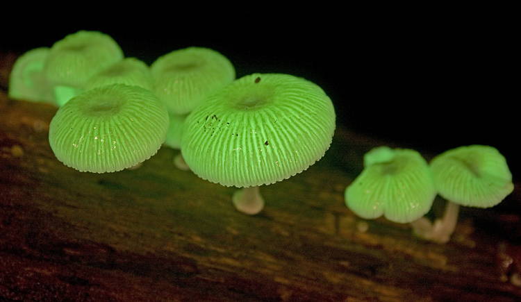
Ans. Glowing mushroom 


Mushroom

                          Image source credit -http://all-that-is-interesting.com/bioluminescence-creatures


3.Bioluminescence is - Light produced by living creatures
4.What is the most common bioluminescent colour in marine life - Blue 
5. Where do most bioluminescent organisms lives -in the deep sea

MCQs.
1.What is the most common source of bioluminescence in surface waters-
a.crustacians                      c.jelly fish 
b.Dinoflagellates                 d. All of the above 
Ans. b
2.What percentage of deep sea creatures are bioluminescent -
a.50 b.45 c. 100 d. 90                                                                  
 Ans. d (90)
3. Which group of living things does not have bioluminescent members?
a. sea squits            c.Flowering plants 
b.bacteria                d.All of the above 
Ans.c
4. Bioluminescence is exhibited by -
a. chlorella  b . chlamydomonas
c. hirudinaria d.ceratium 
Ans.d


Thank you for visiting my blog.Please feel free to share your comment on this
article ,Please subscribe and share the articles to get more such articles.