Thursday 30 March 2017

DOUBLE IMMUNO DIFFUSION

Double immuno diffusion means both the antigens and antibodies diffuse towards each other and form a precipitin line .Ab and Ag placed in separate, opposing wells inside of agarose gel. Solutions diffuse toward one another. A precipitation line is formed at the zone of equivalence where diffusing Ab and Ag forms complex.


Thank you for visiting my video tutorial/ blog.Please feel free to share your comment on this

article ,Please subscribe and share the articles to get more such articles.

Monday 20 March 2017

PRECIPITATION REACTION


Ag is loaded into single well of gel. Gel has been impregnated with specific Ab solution. Ag will diffuse out from well in radial fashion. Diameter of immunoprecipitin ring is proportional o the concentration of the  Ag present.

MCQs.
1.When combining aqueous solutions of soluble compounds, a precipitate is formed where -
 A. a solvent is insoluble
B. a reactant is insoluble
C. a product is insoluble
D. None of these answers is correct. 
Ans. c

2.In agglutination reactions, the antigen is a………in precipitation reactions, the antigen is a……………
a. whole cell/soluble molecule
b. Soluble molecule/whole cell
c. Bacterium/virus
d. Protein/carbohydrates


Ans.a

3.Which of the following serological technique is used to quantify antigen -

A)Enzyme-linked immunosorbent assay
B)Immunoprecipitation

C)Single radial immunodiffusion (RID) assay
D)Immunoelectrophoresis

Ans. c


4.The quantitative technique where a negatively charged antigen is electrophoresed into a gel containing antibody is-

a.RDI (Radial immuno duffusion)
b.Mancini method
c.Double diffusion 
d.Both a and b

Ans.d 

Thank you for visiting my video tutorial/ blog.Please feel free to share your comment on this


article ,Please subscribe and share the articles to get more such articles.

Thursday 16 March 2017

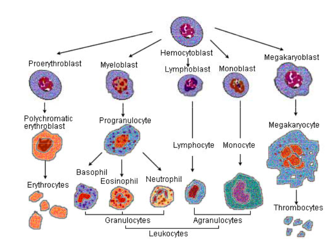
OVERVIEW OF STEM CELL (HAEMOCYTOBLAST) IN HAEMATOPOIESIS

 HAEMATOPOIESIS- Formation of blood cells.

Location-Bone marrow (centre part of the bone when you take a cross section)
Hepatopoitic stem cells -----Progenitor cells(myeloid and lymphoid)-------Precaurser cells (Blast suffix) like  proerythroblast ,myeloblast.monoblast,lymhoblast and megakaryoblast -------Formed elements(RBC,WBC.Platelets).

Platelets or thrombocytes are the progenetors of megakaryocytes.
Granulocytes are precursors are myeloblast(unipotent ) .
Monocyte -Precursor is monoblast (which is found in bone marrow .They mature into monocytes which in turn develop into macrophages .


Primitive hematopoietic stem cells are progenitors for the primitive hematopoietic cells of the Yolk Sac that serve as the source for primitive erythrocytes, megakarocytes and macrophages. These progenitors are committed to a single lineage and appear first in 5th month development.Hematopoiesis originates in the early yolk sac but as embryogenesis proceeds this function is taken over by the fetal liver and spleen, then later by the bone marrow where it continues throughout life.
Erythrocytes and Leukocytes                                             Image source credit -https://www.wikipremed.com/image.php?                                                                     img=040706_68zzzz373750_Illu_blood_cell_lineage_68.jpg&image_id=373750
MCQs.
1.In adults haematopoiesis mainly
occurs in which of the fol¬lowing
A. Long bones B. Liver
C. Spleen D. Flat bones                                        
   Ans.D
2.During foetal life, haematopoiesis commences in the bone marrow by: 
A. 2nd to 3rd month 
B. 4th to 5th month 
C. 6th to 7th month
D. 7th to 8th month
Ans. B
3.Where does haematopoiesis take place?
a) Lungs
b) Pancreas

c) Liver

d) Bone marrow.
Ans. d

4.Which of the following is the earliest site of hematopoiesis in the embryo:
  Bone marrow
  Liver
  Spleen
  Yolk sac
Ans.D
5.Megakaryocytes give rise to-
a.RBC
b.WBC
c.Thrombocytes(Platelets)
d. None of the above
Ans. c
Thank you for visiting my video tutorial/ blog.Please feel free to share your comment on this

article ,Please subscribe and share the articles to get more such articles.

Tuesday 14 March 2017

MONOCLONAL ANTIBODY PRODUCTION BY HYBRIDOMA TECHNOLOGY

A hybridoma, is produced by the injection of a specific antigen into a mouse, procuring the antigen-specific plasma cells (antibody-producing cell) from the mouse and the subsequent fusion of this cell with a cancerous immune cell called a myeloma cell. 
The hybrid cell, which is thus produced, can be cloned to produce many identical daughter clones. These daughter clones then secrete the immune cell product. Since these antibodies come from only one type of cell (the hybridoma cell) they are called monoclonal antibodies. The advantage of this process is that it can combine the qualities of the two different types of cells; the ability to grow continually, and to produce large amounts of pure antibody. 
HAT medium (Hypoxanthine Aminopetrin Thymidine) is used for preparation of monoclonal antibodies. 
Laboratory animals (eg. mice) are first exposed to an antigen to which we are interested in isolating an antibody against. Once isolated from the mammal, the B cells are fused with immortalized myeloma cells - which lack the HGPRT(hypoxanthine-guanine phosphoribosyltransferase) gene - using polyethylene glycol or the Sendai virus. 
Fused cells are incubated in the HAT (Hypoxanthine Aminopetrin Thymidine) medium. Aminopterin in the myeloma cells die, as they cannot produce nucleotides by the de novo or salvage medium blocks the pathway that allows for nucleotide synthesis.


Note-Because of their high specificity, monoclonal antibodies are useful in diagnosing microbial diseases.

MCQs.
1.  In hybridoma technology ,the myeloma cells used lack 
a.HGPRTase
b.the ability to prduce Ig
c.both HGPRT ase and ability to produce Ig
d.thymidine kinase 

ans. c
2.Monoclonal antibodies are -
a.heterogenous antibodies
b.Homogenos antibodies produced from single close of plasma cells
c.Both a and b 
d.None of these

ans.b

3.Hybridoma technology was developed by -
a.Beadle and tatum
b.Francis crick
c.Jacob and monad
d.kohler and milstein

Ans.d

4.
In monoclonal antibody technology, tumor cells that can replicate endlessly are fused with mammalian cells that produce an antibody. The result of this cell fusion is a
A.hybridoma
B.myeloma
C.natural killer cell
D.lymphoblast
Ans. A
5.Hybridomas are produced by combining with-
a.B cell and Myeloma cells 
b.B cell with particular epitopes
c.Both a and b 
d.None of the above
ans.a
6.
All of the following are needed to produce monoclonal antibodies except-
a. Animal to inject
b. Polyvalent antigen
c.PEG
d.None of the above

Ans.b (Polyvalent antigen is not used. A single specific antigen with one epitope is used.)
7.Monoclonal antibodies (mAb) can be potentially used as therapeutic agents. The major advantage is that they can specifically target aberrant cells. However, there is a practical difficulty. Monoclonals are raised in mouse and therefore it is expected that an immune reaction will
develop if these are injected into humans. It is therefore necessary to 'humanize' monoclonal antibody by
1. expressing the genes for the mAb in cultured human cells and isolating the mAb from these cells
2. replacing the Fv region of a mAb with one derived from a human IgG
3. replacing CL and CH regions of the Mab with that obtained from a human IgG.
4. taking a human IgG and replacing the CDRs by those derived from the mouse mAb  (csir june 2011)


Thank you for visiting my video tutorial/ blog.Please feel free to share your comment on this

article ,Please subscribe and share the articles to get more such articles.

Friday 10 March 2017

Agglutination Reaction

asically direct coombs tests is for antibodies against host RBC's, leading to agglutination. With indirect coombs,  we have to look  for antibodies against non-host RBC's (e.g. anti-Rh antibodies), so need to  put the patient's plasma in with other, non-host RBC's and look for agglutination.

People with type A blood will have the A antigen on the surface of their red cells (as shown in the video in a table format) As a result, anti-A antibodies will not be produced by them because they would cause the destruction of their own blood.  However, if B type blood is injected into their systems, anti-B antibodies in their plasma will recognize it as alien and burst or agglutinate the introduced red cells. 
1.
 Agglutinogen in a viral hemagglutination test-
a.Antibodies
b.Antigens
c.WBC
d.RBC

Ans.d

2.
What type of immunologic reaction occurs when specific antibodies are mixed with whole cell antigens?
a.Agglutination This is the correct answer.
b.Precipitation
c.Immunodiffusion
d.complement fixation
Ans. a


Note .The main difference between agglutination and precipitation is the size of antigens.  For precipitation, antigens are soluble molecules, and for agglutination, antigens are large, easily sedimented particles.


Procedure
1. Label three slides 1,2,3.
2.  Wipe your left ring finger with an alcohol wiper and let it air dry.
3. Take a sterile lancet and puncture your fingertip.  
4. Place a small drop of blood on each of three microscope slides.  
5. On the 1 st. slide, place a 20 ml of anti-A antiserum.  Place anti-B on the 2nd. slide and anti-a and anti-b on the 3rd. slide. Mix the antisera in with the blood using a separate toothpick for each slide.  
6. After several minutes, observe agglutination and determine your blood type. So, 
If your blood cells stick together when mixed with:
  • Anti-A serum, you have type A blood
  • Anti-B serum, you have type B blood
  • Both anti-A and anti-B serums, you have type AB blood

If your blood cells do not stick together when anti-A and anti-B are added, you have type O blood.


Test yourself!

1.

Agglutination in all three  indicates that all three antigens are present in the blood: A, B and Rh. Accordingly, the blood type is AB Rh+.
2.

Agglutination in 1st  containg A antibodies and second containing B antibodies. Accordingly, the blood type is AB Rh-.

3.

Agglutination in the test tube with Rh antibodies indicates that the blood has got Rh antigens but no A nor B antigens.
The blood type is O Rh+.

Together, the ABO and Rh grouping systems yield your complete blood type. There are eight possible types: O+, O-, A+, A-, B+, B-, AB+, and AB-.

Thank you for visiting my video tutorial/blog.Please feel free to share your comment on this

article ,Please subscribe and share the articles to get more such articles.


Wednesday 8 March 2017

PRIMARY AND SECONDARY IMMUNE RESPONSE


Primary                                          secondary immune   response             immune                                                              response
Clonal selection refers to the binding by antigen of lymphocytes with a complementary receptor; this occurs in both primary and secondary immune responses.

1.Secondary immune response is generated due to (June 2006) 
a) naive B cells 
b) Memory cells 
c) naive T cells 
d) NK cells
Ans.b
2.Which among following is responsible for humoral immunity?---
A) B-lymphocytes
B) T-lymphocytes
C) L-lymphocytes
D) P-lymphocytes
Ans. A

3.Which class of immunoglobulins will increase in case
 of a chronic infection?
 1. IgA
 2. IgG
 3. IgM
 4. IgE
Ans.2


Thank you for visiting my video tutorial/blog.Please feel free to share your comment on this
article ,Please subscribe and share the articles to get more such articles.

Tuesday 7 March 2017

Overview of innate and adaptive immunity


1. Which of the following does not protect body surfaces:
  Skin.
  Mucus.
  Gastric acid.
  Salivary amylase
Ans.D  The enzyme splits starch and is of importance for digestion but not for protection.

2. Lysozyme:

 A.  Activates complement.
 B  Is a proteolytic enzyme.
C Splits peptidoglycan.
  Is released by mast cells.
Ans. D Lysozyme splits the peptidoglycan of bacterial cell walls.

3. Plasma cells:
  Have a thin layer of cytoplasm
  Are derived from T-cells
  Develop into B-cells
 D  Have a highly developed rough endoplasmic reticulum
Ans. D Plasma cells secrete large amounts of antibody and therefore require well developed ER in order to make sufficient amounts of this protein.

4.A plasma cell secretes:
  Antibody of a single specificity related to that on the surface of the parent B-cell
  Antibody of two antigen specificities
  The antigen it recognizes
  Many different types of antibo
Ans. A  A single plasma cell secretes millions of molecules of antibody but they are all identical and will be related to the single specificity of antibody used as the antigen receptor on the B-lymphocyte from which the plasma cell was derived.


Thank you for visiting my video tutorial /blog.Please feel free to share your comment on this

article ,Please subscribe and share the articles to get more such articles.

Monday 6 March 2017

TOLL - LIKE RECEPTORS


1.What is the name for the cytoplasmic domain of a TLR receptor?
Ans. Toll-IL1-R, or TIR domain

2.The TLR9 pattern recognition receptor recognises:
  CpG motifs.
  Gram +ve peptidoglycan.
  Mycobacterial lipoarabinomannan.
  Gram –ve LPS.  
Correct.answer -A Toll-like receptor 9 recognition of these microbial dinucleotide sequences leads to activation of the NFkB transcription factor.

3.Toll-like receptor 4 is associated with responsiveness to LPS, an endotoxin that causes lethal endotoxic shock. The mice deficient In Toll-like receptor 4 and BALB/c mice were injected with Escherichia coli. In addition, some BALB/b mice were also injected with the same bacteria alone
or with anti-interleukin-10 (IL-10) antibody. The mice resistant to the lethal effect of the bacteria were:
1. BALB/b mice receiving the bacteria.
2. BALB/b mice receiving the bacteria and the anti-IL-10 antibody
3. Mice deficient In Toll-like receptor
4. BALB/c mice receiving the bacteria.


Thank you for visiting my video tutorial/blog.Please feel free to share your comment on this
article ,Please subscribe and share the articles to get more such articles.

Thursday 2 March 2017

JAK-STAT PATHWAY


1.The following hormone act via JAK-STAT pathway except - 
a. GH  b. TSH c. PRL d. IL-6

Ans.b


2.GH produces what other hormone, through the JAK-Stat pathway?
A.  Endothelial growth factor
B. Epithelial growth factor 
C. Insulin-like growth factor


Thank you for visiting my video tutorial/ blog.Please feel free to share your comment on this
article ,Please subscribe and share the articles to get more such articles.