Monday, 19 September 2016

CHECKPOINTS , CYCLINS AND CDK( CYCLIN DEPENDENT KINASE)

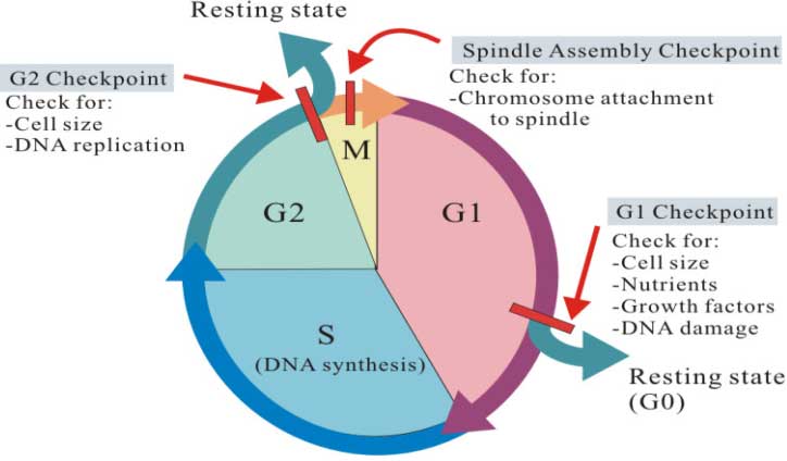
Cell cycle checkpoints-Cell cycle is a sequential step that taking place in a cell leading to the accurate duplication of genetic materials (DNA).  Checkpoints halt the progress of cell cycle if
 (1) any of the chromosomal DNA is damaged
 (2)  DNA replication during S phase or chromosome alignment during M phase, have not been properly completed.  
 Cell cycle checkpoints functions:
  •  The nuclear genome is intact (without any mutation)
  •  The conditions are appropriate for a cell to divide (enough nutrients is there for the daughter cells)
  •   Genetic material is replicated only once in a cell cycle
  •  No mutations occurred in the replicated chromosomes.If mutations are occurred, these mutations will be rectified by DNA repair system
  •  Chromosomes are correctly oriented in the metaphase plate
  •  All chromosomes are correctly attached to the spindle fibres.
The cyclins are so named because their amount varies throughout the cell cycle. To be active, the cyclin dependent kinases (cdks) controlling the cell cycle must bind to a specific cyclin. Cyclins fluctuate during cell cycle.
Different types of checkpoints in cell cycle:


                            Image source credit -http://gleesonbiology.pbworks.com/w/page/7537859/C9

There are three checkpoints in a cell cycle.

(1). G1 checkpoint (restriction checkpoint)
(2). G2 checkpoint (G2-M DNA Damage Checkpoint)
(3). Metaphase (M)-checkpoint (Spindle assembly checkpoint)
(1). G1 checkpoint:
G1 checkpoint(restriction point). operates at the end of G1 phase of cell cycle. It checks whether the conditions are favorable for the cell to divide. It also checks the DNA for any damage before it is going for a cycle of DNA replication in the next phase (S phase). If DNA damage is detected, checkpoint proteins will prevent the formation of active cyclin/cdk complexes. Inhibition of cyclin/cdk complex formation stops the progression of the cell cycle. The cells are then direct the DNA repair mechanism to rectify the DNA damage. If the environmental conditions are not good, the cell may enter into G0 phase. In yeast cells, G1 checkpoint is also called as start point.
(2). G2 checkpoint
G2 operates at the end of G2 phase. ( G2-M DNA damage checkpoint.) G2 checkpoint checks the DNA for any damage that might be occurred during the DNA replication in the previous cell cycle phase (S phase). G2 checkpoint also ensures that the entire DNA has been replicated completely. Apart from this, G2 checkpoint monitors the levels of proteins and growth factors that are needed in the next phase (M phase) of cell cycle. If any of the above factors are not satisfactory, the G2 check point hold the cells at G2 phase and initiate machineries to rectify the problems.  

(3). Metaphase (M) checkpoint (spindle assembly checkpoint)
Metaphase checkpoint is also called as spindle assembly checkpoint. It operates at the end of M phase. Metaphase checkpoint senses the integrity of the spindle apparatus in the cell. Spindle apparatus is involved in sorting of chromosomes during cell division. Correct orientation of chromosomes in the metaphase plate of cell is very essential for the proper segregation of chromosomes. If chromosomes are not correctly attached to the spindle apparatus, the metaphase checkpoint will stop the cell cycle. Thus, M checkpoint prevents cells from incorrectly sorting their chromosomes during division.So, M-Cdk inhibits mitosis .
 Importance of cell cycle checkpoints-
  •  They can induce apoptosis (programmed cell death) .
  •  Cell cycle checkpoint ensure only one round replication of DNA per cell cycle.
  • Almost all cancers are due to the improper functioning of either one or many proteins involved in cell cycle regulation. (Eg. P53 – guardian of genome, a tumor suppressor gene).
Q.1 Tyr-15 is dephosphorylatedby ? 
a. cdc25
 b. cdc2 
c. Both
 d. Cdk1
And. A
Q2.When cell has stalled DNA replication fork, which checkpoint should be predominantly activated?

A)M
B)Both G¹M and M
C)G¹/S
D)G²/M
Ans.D

Thank you for visiting my blog.Please feel free to share your comment on this article ,Please subscribe and share the articles to get more such articles.

Sunday, 18 September 2016

Haemoglobin

Haemoglobin-(Hb )( Respiratory pigment )

It is a globular protein present in RBC which helps in transport oxygen by the help of histidine  . It is a kind of quaternary protein with 4 polypeptide chains (2 alpha and 2 beta ). The heme unit is a ring of atoms which carry an iron atom .(Fe 2+).
Heme binds o2 reversibly .
Co binds to heme more strongly than o2.
Haemoglobin has 2 states-
1.Oxyhaemoglobin (o2 bound 4 nos. )
2.Deoxy- haemoglobin (No O2 bound )

It is found in 2  major conformation forms -
1. R- form (Relaxed form )
-Fewer  interactions ,more flexible
-Higher affinity for O2.

2. T-form (Tense form/ taut stage )

-More interactions ,more stable
-Lower affinity for O2.

Sickle cell disease is a genetic alteration that causes some hemoglobin molecules to be defective (the defective hemoglobin is termed hemoglobin S). 

The iron atom in heme binds to the 4 nitrogen atoms in the centre of the porphyrin ring. Cooperative binding property( uptake of one ligand influences the affinities of the remaining unfilled binding sites. ) Haemoglobin is allosterically inhibited by CO2, protons( which is released from carbonic acid dissociation ) and the body needs oxygen .


 Image source credit -http://cikgurozaini.blogspot.in/2011/09/carbon-dioxide-transport.html


 When oxygen is bound to the first subunit of hemoglobin it change the quaternary structure of the protein this  helps subsequent molecule of oxygen to bind to the next subunit.  This phenomenon is called an allosteric (through space interaction).Iron atom is attached to the proximal histidine, this causes the local helix to move also.


Deoxy Hb   -------  Oxy Hb


Image source credit -http://www.chemistry.wustl.edu/~edudev/LabTutorials/Hemoglobin/MetalComplexinBlood.html

Haemoglobin dissociation curve is S-shaped or sigmoidal.
when Hb combines to one Oxygen, its shape distorts slightly, and that makes it easier for the second Oxygen and so on upto 4th o2 molecule .Fourth can't combine with any more Oxygen molecules since 4 is the maximum number of O2 molecules that can combine with one molecule of Haemoglobin. This diagram reflects the idea:
Dissociation curves

                              http://www.s-cool.co.uk/a-level/biology/transport/revise-it/blog

Q1. When oxygen haemoglobin curve shift to left it represents -(2008 DEC)
a.Decrease in pH
b.Decrese in Co2 level 
c.Rise in concentration of 2,3 BPG
d.more affinity for oxygen 
Ans.  c

Q2.The major role of 2,3 BPG formed during glycolysis in RBC is for haemoglobin is -
a.Increasing affinity for oxygen
b.Decreasing affinity for oxygen
c.Increasing affinity for Co2
d.Decreasing affinity for Co2
Ans. b

Thank you for visiting my blog.Please feel free to share your comment on this article ,Please subscribe and share the articles to get more such articles.

Monday, 12 September 2016

CRYOPRESERVATION

It is a process of preservation of  cells or whole tissues ,  which are preserved by cooling the temperature to low- subzero .(-196c  ) the boiling point of liquid nitrogen . in this all biological process are suspended and material does not decompose .Cryopreserved cells are fragile it should be handle carefully . 


Advantage of freezing cells-
1. Reduced risk of Genetic change .
2.Reduced risk of Senescence leading to extinction of cell line. 
3.Reduced risk of Transformation to tumor related properties. 
4.Reduced risk of Contamination 
5.Saving reagents time 
6.Prevent Cross contamination by other cell line.

Process of cryopreservation -
1.The selection of plant species or tissues with particular  morphological and physiological characters . ( meristems, embryos, endosperms, ovules, seeds, cultured plant cells, protoplasts, calluses.) Among these, meristematic cells and suspension cell cultures, in the late lag phase or log phase are most suitable.

2.Cryoprotectants are the compounds that can prevent the damage caused to cells by freezing .The freezing point of water are reduced by the presence of cryoprotectant. As a result, the ice crystal formation is retarded during the process of cryopreservation.
Cryoprotectants  are dimethyl sulfoxide (DMSO), glycerol, ethylene, propylene, sucrose, mannose, glucose, proline and acetamide. Among these, DMSO, sucrose and glycerol are most widely used. 


3.The sensitivity of the cells to low temperature is variable and largely depends on the plant species.
4.The frozen cells/tissues are kept for storage at temperatures in the range of -70 to -196°C. Storage is ideally done in liquid nitrogen refrigerator — at 1 50°C in the vapour phase, or at -196°C in the liquid phase to maintain viability .For long term storage, temperature at -196°C in liquid nitrogen is ideal. A regular and constant supply of liquid nitrogen to the liquid nitrogen refrigerator is essential. 
5.Thawing is usually carried out by plunging the frozen samples in ampoules into a warm water (temperature 37-45°C) bath with vigorous swirling. By this approach, rapid thawing (at the rate of 500- 750°C min-1) occurs, and this protects the cells from the damaging effects ice crystal formation.
6.Thawed germplasm is washed several times to remove cryoprotectants and  then re-cultured in a fresh medium following standard procedures. 
7.The viability/survival of the frozen cells can be measured at any stage of cryopreservation or after thawing or re-culture.The techniques employed to determine viability of cryopreserved cells are the same as used for cell cultures .Staining techniques using triphenyl tetrazolium chloride (TTC), Evan’s blue and fluorescein diacetate (FDA) are commonly used.
8.Regeneration - For appropriate plant growth and regeneration thr cryopreserved cells should  be carefully nursed, and grown. Addition of certain growth regulators also help in regeneration.

Thank you for visiting my blog.Please feel free to share your comment on this article ,Please subscribe and share the articles to get more such articles.

Saturday, 10 September 2016

LINES & SINES

LINES- (long interspersed nuclear element  )  Autonomous transposons these are foreign sequences that has been inserted into the chromosome. Comprises 21% of the human genome.These L1 elements are DNA sequences that range in length from a few hundred to as many as 9,000 base pairs.
  • The functional L1 elements are about 6,500 bp in length and encode three proteins, including
    • an endonuclease that cuts DNA and a
    • reverse transcriptase that makes a DNA copy of an RNA transcript.
  • Active L1  element is about 6kb long and consists of 2 ORFs ( ORF1 &ORF2), 5' UTR ( untranslated region) ,3' UTR contains poly A signal.
  • ORF1 binds to L1 m-RNA , ORF2 contains reverse transcription activity  and endonuclease domain .
5'UTR---------------O---------------------------------3' AAAAAAA ( m-RNA )

                             ORF1                    ORF2

SINEs- ( short  interspersed nuclear element )Non -autonomous                
 DNA sequences (100–400 base pairs) that represent reverse-transcribed RNA molecules originally transcribed by RNA polymerase III.The most abundant SINEs are the Alu elements. There are over one million copies in the human genome (representing 9% of our total DNA).
Alu elements consist of a sequence averaging 260 base pairs that contains a site that is recognized by the restriction enzyme AluI. 


Thank you for visiting my blog.Please feel free to share your comment on this article ,Please subscribe and share the articles to get more such articles.

Thursday, 8 September 2016

Rolling circle mechanism

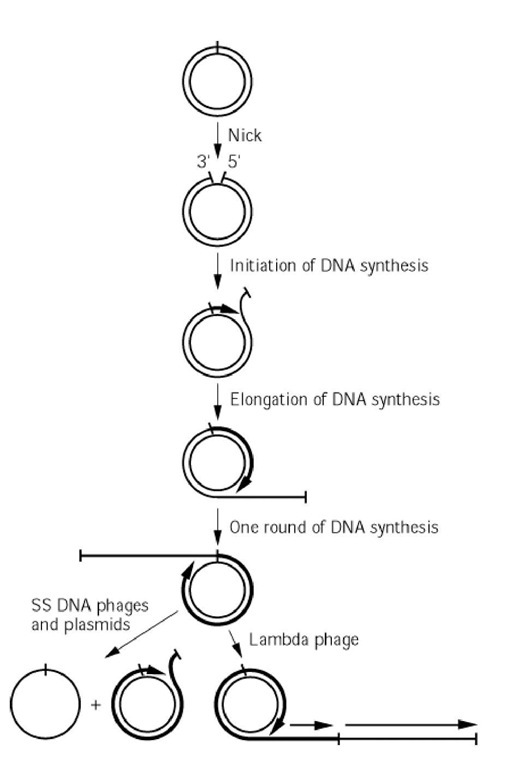
Rolling circle mechanism  is an alternative method used by bacteriophages and viruses for the transfer of plasmid DNA.

One strand of DNA is nicked to provide the 3' OH group. DNA synthesis begins at the 3' end of the broken strand , the other strand is used as a template . SSB and helicase create a replication fork and nicked strand (5' end of the broken strand is displaced). Replication is initiated by a break in one of the nucleotide strands.

Cleavage releases a single -stranded linear DNA and a double stranded circular DNA. 

The linear DNA may circularize and serve as a template for synthesis of a complementary strand . dNTPs are added to the 3'OH group to create the leading strand .

Rep proteins are encoded by RC plasmids that contains specific domains which are involved in origin binding and nicking activities. 

The products of rolling -circle replication are multiple circular DNA molecules .


                                              Image result for rolling circle mode of replication
Image source-http://what-when-how.com/molecular-biology/rolling-circle-dna-replication-molecular-biology/
Thank you for visiting my blog.Please feel free to share your comment on this article ,Please subscribe and share the articles to get more such articles.

G0-phase

Many times a cell will leave the cell cycle ,temporarily or permantently. It exists the cycle at G1 and enters a stage designated G0.(resting phase) It is also called as quiscent stage where the cell is neither dividing nor preparing to divide.

Often G0 cells are terminally differentiated they will never re-enter the cell cycle but instead will carry out their function in the organism untill they die.

Hepatocytes in the normal liver are quiesent (G0 stage ).

G0  represents not simply the absence of signals for mitosis but an active repression of the genes needed for mitosis. Cancer cells cannot enter Go stage as they are designed to repeat the cell cycle indefinitely .

 Some types of cells, such as nerve and heart muscle cells, neurons become quiescent when they reach maturity but continue to perform their main functions for the rest of the organism's life. Multinucleated muscle cells that do not undergo cytokinesis are also often considered to be in the G0 stage.  

During the G0 phase, the cell cycle machinery is dismantled and cyclins and cyclin-dependent kinases disappear. 


                              Image source-https://14milema.files.wordpress.com/2010/10/cellcycle2.png


The importance of G0 phase - During cell division when the cell get a signal that there are enough of cells  or there is a mutation in the cell that need to be fixed before they differentiate fully or the cell during its division gets damage then it is signalled to rest, and it is this phase that cell leave the cycle and quit dividing. It could be temporary resting period (so when cell again required, or damaged got fixed) then the cells will again join the cycle and continue to divide again or it can also be permanent for example neurons, which are resting in G0 phase and never divides again.
1.
Terminally differentiated cells are most often found in which phase of the cell cycle?
A. G0
B. G1
C. G2
D. M
Ans. A
Thank you for visiting my blog.Please feel free to share your comment on this article ,Please subscribe and share the articles to get more such articles. 

Wednesday, 7 September 2016

Catabolite repression

In case of catabolism of carbohydrates catabolic breakdown  of glucose which is a simple carbohydrate prevents activation (repression) of the lac operon by lactose, so this effect was originally called catabolite repression a positive control of lac-operon. It is found in various bacteria and other micro-organisms. 

In a culture medium of bacteria if we have both glucose and lactose . Suppose the concentration of lactose is more as in comparision to glucose. Then, glucose represses the activity of lactose . It will not allow the bacterial cells to uptake lactose first  unless untill all the glucose molecules get metabolized or depleted.
It is also called as Glucose effect  as in this case the glucose is the repressor .


Process-
Permease protein allows to pass the lactose inside the cell and the glucose by glucose transpoters. Glucose keep phosphotransferase enzyme in active condition which is present in the the bacterial cell. .(Switch off mechanism) it looses phosphate group and it block  adenyl cyclase enzyme production(which produces cAMP). Transcription process is not possible due to lack of beta- galactosidase and permease .

Furthermore, when glucose levels are low the phosphorylated form of phosphotransferase accumulates and consequently activates the enzyme adenylyl cyclase, which will produce high levels of cAMP. cAMP binds to catabolite activator protein (CAP) and together they will bind to a promoter sequence on the lac operon. 

Thank you for visiting my blog.Please feel free to share your comment on this article ,Please subscribe and share the articles to get more such articles.
Tuesday, 6 September 2016

Richmond-lang effect

Richmond- lang effect-

This effect is discovered by Richmond & Lang in 1967. Acc. to this effect the delay of senescene of leaves and other parts takes place by the help of plant growth regulator cytokinin.

Leaf senescence is a developmental process actively initiated as part of an age-dependent genetic program or in response to environmental stress which leads to apoptosis.

Retardation of senescence leads to retard the rate of degradation of chlorophyll,keeping them productive longer. It  has an effect on protein synthesis and mobilising nutrients.

Cause- It was found that increased cell-wall invertase activity which occurs in response to cytokinin is both necessary and sufficient for the inhibition of senescence.


Thank you for visiting my blog.Please feel free to share your comment on this article ,Please subscribe and share the articles to get more such articles.

Wednesday, 31 August 2016

Leghaemoglobin protein

Leghaemoglobin protein- In case of nitrogen-fixing root nodules of leguminous plants which belongs to family fabaceae nodule formation takes place.

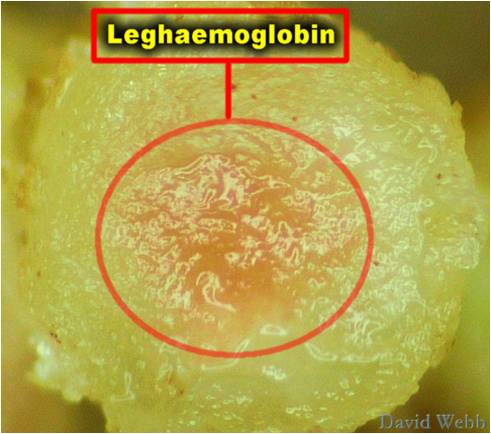
Nodules contain an oxygen binding heme protein called leg-haemoglobin . 

Location -This protein is present in cytoplasm of infected nodule cells at high concentration (700uM). 

It gives the nodule a pink colour apperance. The host plant produces the globin portion of the leg haemoglobin in response to infection by rhizobia bacteria. The bacterial symbiont produces the heme portion. 

Properties-

  • Leg haemoglobin has a high affinity for oxygen about 10 times higher than B-chain of human haemoglobin.
  • It stores enough oxygen to support nodule respiration for a few seconds.
  • Leghemoglobin buffers the concentration of free oxygen in the cytoplasm of infected plant cells to ensure the proper function of root nodules.


Picture

                                Image source-http://biology4isc.weebly.com/3-mineral-nutrition.html


Thank you for visiting my blog.Please feel free to share your comment on this article ,Please subscribe and share the articles to get more such articles.

Monday, 29 August 2016

Hydropathy index

Hydropathy index-  

It is related to amino acids. It represents the hydrophobic properties of amino acid side chains.The larger the number in the scale the more hydrophobic the amino acid .


HYDROPATHY SCALES (a measure of hydrophobicity) -- a way of quantitating whether a 20 amino acid segment might be hydrophobic enough to be a transmembrane helix. More hydropathy index means more energy is required to stabilize in the aquous environment .


  • More + numbers indicate more hydrophobic R groups;
  •  more - numbers indicate more hydrophilic R groups.
The free energy change accompanying movement of amino acid side chain from hydrphobic solvent into water .

Charged (polar )-Exergonic Aromatic and Aliphatic-Endergonic 
Hydropathy plots (plot of hydropathy index vs. residue number where the residue number is actually the average hydropathy value for a "sliding window" of, say, 7 residues, moving down the protein from N to C-terminus -- for example, average values for residues 1-7, then for 2-8, then 3-9, etc. are plotted.
  • Depends on the hydropathy scale being used (i.e., what nonpolar solvent was used for the partitioning experiments to determine relative solubilities in H2O vs. nonpolar solvent); zero is always the midpoint of being neither hydrophilic or hydrophobic, and negative values are more hydrophilic, while positive values are more hydrophobic.


                                           Image source credit- http://slideplayer.com/slide/7360690/


Thank you for visiting my blog.Please feel free to share your comment on this article ,Please subscribe and share the articles to get more such articles.