Thursday, 21 July 2016

Entner -Doudoroff Pathway

Entner -Doudoroff Pathway- (By Michael Doudoroff and Nanthan Entner )

It is an alternative pathway for production of puruvate from glucose. It is different from glycolysis pathway. It uses many enzymes from both Pentose phosphate pathway and glycolysis.

It occurs in prokaryotes specially in the Gram (-)ve bacteria for example pseudomonas aeruginosa, Azotobacter,Rhizobium etc. It absent in eukaryotes.

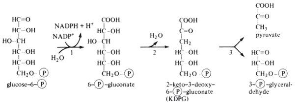
In this pathway glucose-p is oxidised to 2-keto -3-deoxy -6-phosphogluconic acid. It is cleaved by 
 2-keto -3-deoxyglucose -phosphate aldolase to pyruvate and glyceraldehyde -3-phosphate .

It thrn oxidised to pyruvate by glycolytic pathway . 2 ATPs are produced by substrate level phosphorylation.


Picture
                                 Image source -http://microbiochem.weebly.com/e


Thank you for visiting my blog.Please feel free to share your comment on this article ,Please subscribe and share the articles to get more such articles.

Tuesday, 12 July 2016

EtBr

EtBr- It is a planar ,aromatic,intercatalytic agent. It is used in molecular biology labs as a fluroscent dye for nucleic acid gel electrophoresis.

It binds non covalently to ds Dna by intercatalyting into the stacked base pairs. It forms close vanderwall contacts with the base pairs of  DNA and interact with hydrophobic interior of DNA molecule. 

One EtBr molecule integrated upto 2.5 bp of DNA and causes the DNA to unwind by 27°C in covalently circular DNA .Hence, it decreses the number of helical turns. When EtBr binds to linear DNA it causes the helical pitch to increase.

Effects-
1.Ethidium Bromide (EtBr) is sometimes added to running buffer during the 
separation of DNA fragments by agarose gel electrophoresis.The mobility of 
DNA was always less in the gels with EtBr.

2.Ethidium bromide possesses UV absorbance maxima at 300 and 360 nm. It 
can absorb energy from nucleotides excited by absorbance of 260 nm radiation.


3.In covalently-closed circular DNA(plasmid), twist (turns)and writhe(supercoiling) are directly related.  With ethidium, the negatively supercoiled plasmid ( E. coli) becomes positively supercoiled, and each additional ethidium incorporated further positively supercoils it, like winding a telephone wire.


                     Image source-http://sandwalk.blogspot.in/2007/07/ethidium-bromide-binds-to-dna.html
1.Ethidium Bromide is used in electrophoresis of DNA fragments because:
[a] It makes the fragments visible under UV light
[b] It makes the DNA fragments more mobile in the gel
[c] It increases the conductivity of electricity through the gel
[d] It helps determine the size of the fragments
Ans.  A

Thank you for visiting my blog.Please feel free to share your comment on this article ,Please subscribe and share the articles to get more such articles.

Monday, 11 July 2016

Edman degradation

Edman degradation -(Pher Edman)

Definition-It is a method to determine the sequence of peptides  and proteins from the N- terminal of a polypeptide chain.
Reagent-  Phenylisothiocyanate

Mechanism-

 1.Under alkaline condition-

  • Phenylisothiocyanite reacts with N-terminal of amino group of polypeptide .
  • The product of the above reaction is phenylthiocarbamyl-peptide derivative (PTC-peptide.)
2. Under acidic condition-
  • In the presence of trifluroacetic acid this product cleaved as a thiazolinone derivative.
  • It then form phenylthiohydantoin which can be easily identified by using chromatography or electrophoresis.


Thank you for visiting my blog.Please feel free to share your comment on this article ,Please subscribe and share the articles to get more such articles.
Molecular chaperones

Molecular chaperones- 

The spontenity folding of protein is not takes place in case of all the proteins. Some proteins need  a class of  other proteins  for a proper folding. The other proteins are called chaperones.

It is present in both prokaryotic and eukaryotic structures. Chaperones were 1st identified in bacteria E.Coli.

1.Hsp 60( 60 kDa) [ GroEL/GroES in E.coli ] Also called chaperonins forms barrel shaped structure.
GroEL (Larger heptameric structure with 14 identical residues )HSP60 in E.coli)binds to many unfolded proteins . Interaction with the substrate is based on hydrophobic interactions between surfaces of the substrate and residues of GroEL that are exposed in its central cavity. ATP and protein cap GroES (Smaller heptameric subunit with 7 identiical subunits) binds.  ATP hydrolysis occur for release of GroES and the protein molecule. A  new unfolded  protein molecule can now bind to GroEL.

                                       
2.Hsp 70 (70 kDa) It is induced by stress i,e heat shock . Both eukaryotic and prokaryotic.Bind with partially folded polypeptides with exposed hydrophobic regions and prevent aggregation.

The E.coli analogs of HSP70 and Hsp40 are Dna k and Dna J respectively.

Dna J binds to unfolded protein and then to Dna k. Dna J stimulates ATP hydrolysis by Dna k. Dna-k Adp binds tightly to the unfolded proteins.
In bacteria the nucleotide exchange factor Grp E stimulates release of ADP.
ATP binds to Dna k and then the protein dissociates .


Note -Chaperones are used to prevent misfolding and aggregation .
It is necessary in crowded intracellular environment to prevent aggregation .

1.-----------------mediate protein folding .
a.Chaperones
b.Ribosomes
c.Protein itself
d.None of the above
Ans. a


Thank you for visiting my blog.Please feel free to share your comments .Please subscribe and follow me in Google + to get notified about new  articles .

Friday, 8 July 2016

Molten globule state

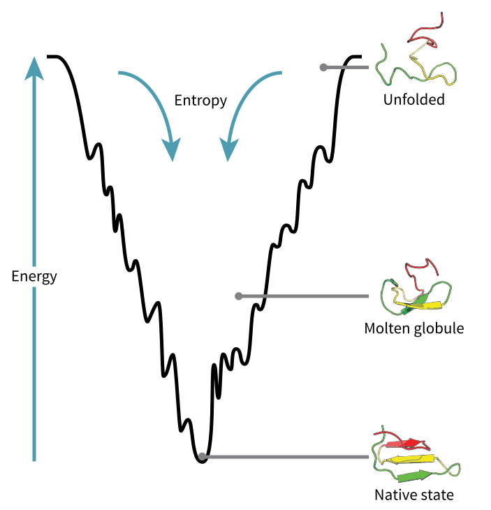
Definition - It is an intermediate stage  in the protein folding pathway .(unfolded protein- molten globule -folded protein)
  
The protein folding pathaway is complicated . There are many models in support of this one of them is the hydrophobic collapsed model (molten globule) . It is called as a collapsed stage as the protein folding is initiated by a spontaneous collapse of the unfolded stage into a globular stage mediated by hydrophobic interactions.

This event is very fast (milliseceond).

Properties-

  • It has most of the secondary structure in the native form.
  • It is less compact.
  • It lacks proper packing interactions in the interior of protein.
  • The interior side remain mobile just like liquids.
  • It can be measured by Circular dichroism (CD) and NMR (nuclear magnetic resonance) .
  • It has a slowly fluctuating tertiary structure .
  • Not specific assembly of secondary structure and the interactions.
  • The initial collapsed state of a folding protein is known as a molten globule. However, a molten globule’s side chains are extensively disordered, its structure fluctuates far more than that of the native protein, and it has only marginal thermodynamic stability. 
                                         

                           Image source-https://en.wikipedia.org/wiki/Hydrophobic_collapse#/media/File:Folding_funnel_schematic.svg


  • Thank you for visiting my blog.Please feel free to share your comment on this article ,Please subscribe and share the articles to get more such articles.

Friday, 17 June 2016

CYBRIDS

 Cybrids- It is also called as cytoplasmic hybrid.  It consist of 2 protoplast one is donor and other is recipient the nuclear content is eliminated forming a normal heterokaryon while its plastome(cytoplasmic genome) or chondriome (mitochondrial genome )content are merged with the recipient. 


  • Cybrids are cells  containing nuclear genome from only one parent but cytoplasm from both the parental species. The process of protoplast fusion resulting in the development of cybrid is known as cybridization.
  • It possess heterozygosity of extrachromosomal genes. It may be single cell or a complete growing plant called cybrid plants.

Applications-

1.Way for those species witch don’t perform to sexual reproduction with each other, so this way provided to facilitate to make a desire species with combination of both species.

2.Production of wide range of genetic variations. 
3.Useful for sterile plants, mitochondrial gene also combined with chloroplast genes of another species.


Thank you for visiting my blog.Please feel free to share your comment on this article ,Please subscribe and share the articles to get more such articles.

Thursday, 9 June 2016

S1 nuclease mapping

S1 nuclease mapping -
    
S1 nuclease is an endonuclease which is  specific for hydrolyzing single-stranded RNA or DNA molecules. (zinc-requiring ) It is more active on DNA then RNA. The protein is thermostable and resistant to several denaturing agents, such as urea, SDS, and formamide . 
m-RNA  is hybridized to ss DNA( radiolabelled with a probe ) that overlaps the start of the target transcript . It  results in DNA-RNA hybrid. The hybrid molecule has single stranded extensions that are degraded by the single stranded specific S1 nuclease . The 3' end of DNA fragment has been removed by using electrophoresis .

                        Image source-http://biocadmin.otago.ac.nz/fmi/xsl/bioc2/learnbitslecture.xsl?-db=BIOC2web.fp7&-lay=Lectures&-recid=4841&-find=

Thank you for visiting my blog.Please feel free to share your comment on this article ,Please subscribe and share the articles to get more such articles.

Tuesday, 7 June 2016

Melting temperature (Tm)


(Melting temp.-Tm)

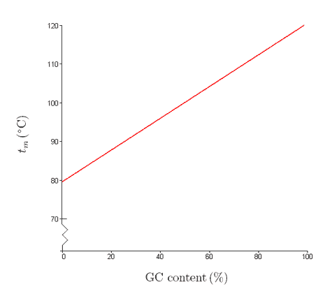
The temp. at which half the strands of DNA are single stranded and half are double stranded. Half of the DNA are denatured . (Tm) It  is characteristics of Dna compostion ( GC content) .The higher the G+C content higher the Tm. 


Image source-http://cbc.arizona.edu/classes/bioc461/Chapter5Part1Notes.htm
Image result for melting temperature of dnaImage result for melting temperature of dna


Image source-http://www.biology.arizona.edu/biomath/tutorials/linear/linearfunctionapplication/dnamelt.html
            
                      Tm directly proportional to G+C content 

Q.What is the maximal tm for a DNA sample that is 500.0 bp long?tm = 64.9 + 0.41(%GC) - (500/length of DNA).

Tm is useful for monitoring your PCR reaction because it lets you distinguish between specific and unspecific amplification. Of course it is easy to see if you have dimer formation if you run your PCR products on a gel, but
with many samples it soon becomes tedious.


You can monitor DNA melting through UV light. The aromatic bases in DNA absorbs UV light (260nm). But when the two strands are hydrogen bonded with each other, the UV light is not absorbed readily. However, when they melt, the bases are exposed and the absorption goes really high. So if you shine UV light onto your sample and raise the temperature of your sample, you will see a sudden increase in absorbency when the DNA melts.

The Tm can be determined experimentally or calculated from simple formula -
   
    Tm=(4x [G+C]) + 2x[A+T]) 

Factirs that affect the Tm -
1.Concentration of DNA.
2.Concentration of ions in the solution ,most notably Mg2+ and K+.
3.DNA sequence
4.Length of Dna.

Thank you for visiting my blog.Please feel free to share your comment on this article ,Please subscribe and share the articles to get more such articles.


Tuesday, 31 May 2016

Blue white screening

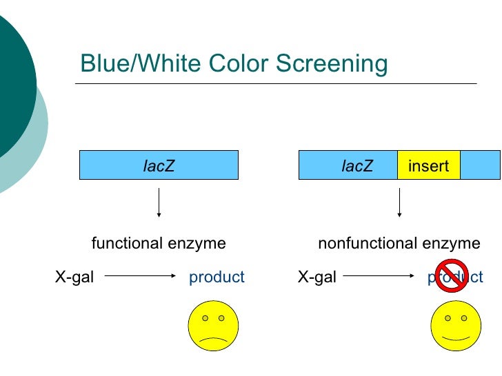
Blue white screening- Foriegn DNA and plasmid Dna are cut with restriction enzyme. Plasmid has gene for lactose hydrolysis lac z gene which codes for enzyme (β-galactosidase). Foriegn dna insert into lacz gene to form the recombinant. The recombinant dna is then transfered into a host cell , which are then grown in the presence of X-gal. 

Result-
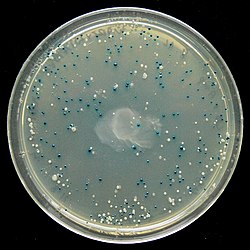
colonies with an insert-containing plasmid have a non-functional β-galactosidase, and remain the whitish-cream color of standard E. coli. On the other hand, intact β-galactosidase ( non recombinant plasmid )produces pigment from x-gal (included in the transformation plate medium), turning the bacterial colony blue.


Blue/White Color Screening lacZ lacZ insert functional enzyme nonfunctional enzyme X-gal product X-gal product


Imagehttps://en.wikipedia.org/wiki/Blue_white_screen


                                                                                                                                                                                                                                                                                                                                                            Imagesourcehttp://www.slideshare.net/tejondaru/blue-white-selectionpresentation

Thank you for visiting my blog.Please feel free to share your comment on this article ,Please subscribe and share the articles to get more such articles.

Linkers and adapters

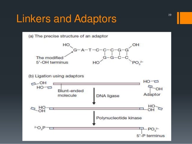
Linkers- Short stretches of dsDNA of known  nucloetide sequence (length 8-14 bp )and have a recognition site for 3-8 restriction enzymes.

Ligated to blunt ends of DNA by ligase.Because of the high concentration of these small molecules present in the reaction, the ligation is efficient when compared with blunt-end ligation of large molecules. The cohesive ends are generated by digesting the DNA with appropriate RE that generates cohesive ends by cleaving in the linkers. The problem with linker is that the sites for the enzyme used to generate cohesive ends may be present in the target DNA fragment. This drawback limits the use of linkers for cloning. 

Adapters are linkers with cohesive ends or a linker digested with RE, before ligation.
It already has one sticky end.The most widely used definition is cut linkers also called as adapters. 

They are not perfectly double stranded non single stranded. By adding adaptors to the ends of a DNA, sequences that are blunt can be converted into cohesive ends and used for cloning .


Sticky ends are desirable for DNA cloning , but sometimes blunt ends are present .Blunt ends will binds to the blunt ends of target to form sticky ends.


Linkers and Adaptors
19

                         Image source-http://www.slideshare.net/drsandeepaiims/gene-cloning-sandeep-jan-2016

Thank you for visiting my blog.Please feel free to share your comment on this article ,Please subscribe and share the articles to get more such articles. 
Monday, 30 May 2016

Bioscience formulae

Bioscience formulae-

1.VP=VG+VE+VGE

2. Crossover value by the square root of the number of individuals (.\/N)

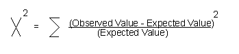
3.Chi-Square Formula-
Degrees of freedom (df) = n-1 where n is the number of 

4.No. of different genotypes-n(n+1)/2,3^n

5.No. of different gametes and phenotype - 2n

6.Recessive homozygous in F2 =1/(2n)2

7.Frequency of recombination-no.of recombinant/Total no. of progeny*100

8.Map distance - No. of recombinant offspring/Total no. of offspring*100

9.Distance betwen 2 genes- No. of single CO+ No. of DCO/Total no. of progeny *100

10.Coincidence- (observed DCO/ expected DCO)

11.Interference=1-(observed DCO/ expected DCO)

12.In tetrad analysis-

a.Map distance= 1/2*No. of SDS asci  / Total no. of asci *100
b.Recombination frequency-NPD+1/2 TT / Total no. of tetrads *100

13.No.of F2 progeny expressing either extreme phenotype- 1/ 4^n

14.Inbredding coefficient= 2pq-H/2pq( population genetics)

15.Ne= 4NmNf /Nm+Nf( Effective population size)

 Nm= no.of male Nf= no. of female

16.dC/dt= -kC^2    dC/dt=rate of renaturation
   
     C0t1/2 = 1/k 

17.p  ( buoyant density )

=1.660 +0.00098 (% GC)g/cm3 

18.Packaging ratio of DNA= Extended length of DNA/ Packing length


19.Dna replication time= base pairs/ no. of bases per sec

20. Size of fragments = 4^n  ,where n= no. of base pairs


21.Time since divergence of two species is given by

(100 - X% sequence similarity) / (% change / years).

22.pH = -log[H+] or pH = -log[H3O+]

23. Kw = 1.0 x 10-14 =  [H3O+] [OH-]  

24.Viability of cryopreserved cells -


25.Determining the no. of genes=1/4 ^n

26.Nt=No+B+I-D-E     I=Immigration  B=Natality rate   D= Mortality rate 

(change in population size at a given time interval t)


Thank you for visiting my  blog. Please share your comments on this

article.  Subscribe and follow me in Google + to get notified with new articles.

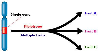
Pleiotrophy

Pleiotrophy - One gene have the ability to produce two or more unrelated phenotypic traits. 
An example is phenylketonuria.
The disease causes mental retardation, reduded hair and pigmentation.
Due to mutation in a single gene phenylalanine to tyrosine.


                            Image source-http://mumtazticloft.com/PigeonGenetics1.asp

Other examples are-
1.Sickle cell anemia -enlarged spleen,muscle pain,low RBC count
2.Albinism- Albinism is also a pleiotropic trait affecting eyesight as well as resulting in a lack of pigmentation in skin, hair, and eyes.
3.Cytokines that have different biological functions.


Thank you for visiting my blog.Please feel free to share your comment on this article ,Please subscribe and share the articles to get more such articles. 
Saturday, 28 May 2016

Flow cytometry

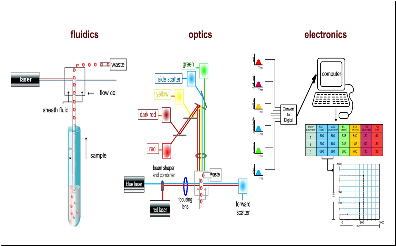
Flow cytometry- It is used to measure the number of cells from a suspension of heterogenous population. It can analyze and sort particles on a one by one basis at rates of 50 000 particles per second. It consist of 3 main components-

1.The flow system
2.The optical system
3.The electronic system

Flow system-  The sample is injected into the centre of a stream of liquid (water or buffer), called the sheath fluid. the particles are randomly distributed.


The optical system-
After hydrodynamic focusing, each particle passes through one or more beams of light(lasers).Light scattering or fluorescence emission (assumed the particle is labeled by a fluorochrome) provides information about the particle’s properties. Fluorescence measurements taken at different wavelengths can provide quantitative and qualitative data about fluorochrome-labeled cell surface receptors or intracellular molecules such as DNA and cytokines. Light is quantified by detectors.The specificity of detection is controlled by optical filters, which block certain wavelengths while transmitting others. Charged droplets are deflected electrostatically by passage through an electrical field.
The electronic system- It analyze statistically by software to report cellular components, size,phenotype .

Applications of Flow Cytometry-

Typical applications of  flow cytometry include-
  • Biomolecular studies
  • Immunology
  • Single cell analysis
  • Medical applications (hematology, genetics)
  • Autofluorescence characterization. 
1.Flow cytometer is used to measure the number of-
A. Cells                    c. Proteins
B. Dna                      d. Rna
Ans.  A (cells) 

Thank you for visiting my blog.Please feel free to share your comment on this article ,Please subscribe and share the articles to get more such articles. 
Friday, 27 May 2016

Promiscuous dna

Promiscuous dna- Endosymbiotic transfer of DNA from cytoplasmic organelles (mitochondia and chloroplast) to the nucleus.  It is supposed that the chloroplasts and mitochondria were once free-living cells that linked up with the embryonic plant cell to form a symbiotic partnership.


                         Image source -http://www.nature.com/scitable/content/organellar-dna-mobility-and-the-genetic-control-14665064

The eukaryotic mitochondria is derived from a proteobacterial endosymbiotic ancestor . Most of the genes that were originally present in this ancestor's genome have been transferred to the nucleus .With only a small number being retained in the organelle (MITOCHONDRIA). Similarly, most of the genes from the cyanobacterial endosymbiont ancestor of the chloroplast were also transferred to the nucleus.

 So, as a result, cytoplasmic organelles are heavily dependent on nuclear genes and import more than 90% of their proteins from the cytoplasm. The dotted arrows indicate how DNA of mitochondrial (blue) and chloroplast (green) origin is still being transferred to the nucleus.Chloroplasts genes have now been found inside plant mitochondria.


Thank you for visiting my blog.Please feel free to share your comment on this article ,Please subscribe and share the articles to get more such articles. 

Thursday, 26 May 2016

Flippase /Floppase /Scramblase

1.Flippase(P-type ATpase) -Transporters that move lipids to the cytoplasmic face of the membrane are commonly called “flippases.  It uses ATP to move the aminophospholipids  such as PS(phosphatidyl serine) and to a lesser extent, phosphatidylethanolamine (PE), from the outer leaflet to the inner leaflet of the plasma membrane against the concentration gradient.

2. Floppases (ABC transporter)- Transport lipids from the cytofacial surface to the opposite side of the membrane are called “floppases” .it uses ATP to transport substrates such as phosphatidylcholine (PC), sphingolipid (SL) and cholesterol against concentration gradients in the opposite direction. 

Movement of phospholipid molecules between the two leaflets that compose a cell's membrane (transverse diffusion, also known as a "flip-flop" transition). 

3.Scramblase-It moves lipids across the bilayer in either direction towards equilibrium. It briefly disrupt random assymetry.


1.Flippase  2.Floppase 3.Scramblase

                             Image source-https://www.studyblue.com/notes/note/n/micro-2200-study-guide-2011-12-kang-/deck/9733592

Thank you for visiting my blog.Please feel free to share your comment on this article ,Please subscribe and share the articles to get more such articles. 
Wednesday, 25 May 2016

Lipid rafts

Lipid rafts - The plasma membrane is composed of a lipid bilayer  inserted various proteins(transmembrane ion-channel and transporter proteins). Many of these proteins are found associated with surface of the membrane which are rich in free cholesterol and sphingolipids (lipids containing the amino acid sphingosine ).

Lipid rafts are more ordered and tightly packed than the surrounding bilayer, but float freely in the membrane bilayer.

Lipid rafts generally contain 3 to 5-fold the amount of cholesterol found in the surrounding bilayer. Also, lipid rafts are enriched in sphingolipids such as sphingomyelin and play important roles in signal transduction .


https://www.researchgate.net/figure/230685775_fig6_Fig-12-Structure-and-formation-of-lipid-rafts-based-on-a-lipid-ordering-disordering

Types of lipid rafts-
1.Caveolae type- Flask shaped invaginations in plasma membrance which is riched in caveolin.
2.Planar lipid rafts- Found in neurons and enriched in flotilin.
3.Glycospingolipid enriched membranes (GEM)
4.Polyphosphoinositol rafts 


Types of lipid rafts
                                      http://www.getmededu.com/plasma-or-cell-membrane.html
FUNCTIONS -
1.Provide stability or rigidity .
2.Helps in signal transduction.
3.In endocytosis it helps.
4.Cholesterol transport .

Thank you for visiting my blog.Please feel free to share your comment on this article ,Please subscribe and share the articles to get more such articles. 

Cloning

 Cloning: (Dolly)

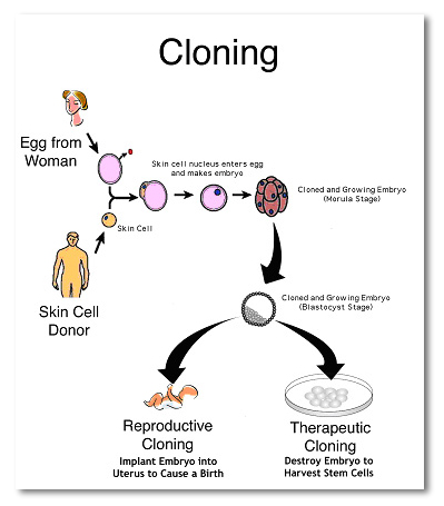
 Definition-Clones are organisms that are exact genetic copies. 
 Every single bit of DNA is identical. It can happen naturally— identical twins are
 just one of many  examples or can be made in the lab.

a) First clone - tadpoles using embryonic cells and nuclear transfer.

b) Why cloning?

  1) cloned animals produce proteins to treat disease. 
  2) for organ transplantation
  3) therapeutic human cloning and stem cells.

c)Types of cloning-
    1.Therapeutic cloning-by the use of stem cells for research.
    2.Reproductive cloning- for cloning human and animals.
                           Image source-http://mrdclassified.weebly.com/cloning-and-gmos.html


 3.Gene cloning-DNA derives from genome of organism and cloned into a cloning
    vector (Plasmid).
                               

Thank you for visiting my blog.Please feel free to share your comment on this article ,Please subscribe and share the articles to get more such articles.