Wednesday, 25 May 2016

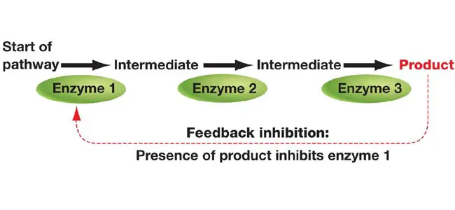
Feed-back inhibition:
Feed-back inhibition: 

  • An allosteric enzymatic activity regulation mechanism in the cell to regulate metabolic pathways with more than one steps and includes more than one enzyme. 
  • Usually the first enzyme in these metabolic pathways will be an allosteric enzyme. Allosteric enzymes have “other sites” for the binding of modulators. Binding of modulators may stimulate or inhibit the activity of allosteric enzyme temporarily for short period of time.
  •  In feed-back inhibition, the end product of the metabolic pathway act as the negative modulator which binds to the allosteric site of the first enzyme and thereby making it unable to bind to the substrate and ultimately halts the metabolic pathway for a period of time until all the end products are consumed. The modulator only binds non-covalently (temporarily) to the allosteric enzyme.
  • Feedback inhibition works by deactivating an enzyme using the product of the reaction the enzyme catalyzes. Enzymes bind to molecules with active sites that are specifically designed to fit with the molecule undergoing the reaction. These enzymes have a second active site for the reaction product to bind to. This causes the enzyme to spatially re-arrange so it can no longer bind to the initial reagent and the reaction stop.
  • Citrate is used for feedback inhibition, as it inhibits phosphofructokinase, an enzyme involved in glycolysis that catalyses formation of fructose 1,6-bisphosphate, a precursor of pyruvate. This prevents a constant high rate of flux when there is an accumulation of citrate and a decrease in substrate for the enzyme.

                                         Q.1Reaction products inhibit catalysis in enzymes by:
a. Covalent binding to the enzyme
b. Altering the enzyme structure
c. Occupying the active site
d. Form a complex with the substrate
Ans: b- Altering the enzyme structure
Q2. A woman patient suffering from thyrotoxicosis shows high level of thyroxine in the blood which is attributed to failure in feed back inhibition in hypothalamic-pituatory-thyroxine circuit. If we further check blood in detail, patient will also show high level of
A: TSH
B: thyroid stimulating IgM
C: TRH
D: parathyroid hormone
Which of the following combination of above statements is correct?
a) A and B
b) B and C                                                                   Ans.B
c) A and C
d) B and D
Q3.Which factor is responsible for inhibition enzymatic process during feed back?
a) Enzymes
b) End product
c) Temperature
d) Substrate                                                                 Ans.B

Q4.Intensity of light increase 20 times, rate of photosynthesis will
(A) increase 
(B) not increase
(C) decrease 
(D) increase till feed back inhibition                                   Ans.D

Q5.The ability of CTP (cytidine triphosphate) to bind to aspartate carbomyltransferase and shut down the synthesis of more CTP is an example of-
1.enzyme repression
2.enzyme induction
3.feed back inhibition   (Ans.)
4.channeling

Q6.Feedback inhibition differs from repression because feedback inhibition
a. is less precise
b. is slower acting
c. stops the action of preexisting enzymes                             Ans. C
d. stops the synthesis of new enzymes

Thank you for visiting my blog.Please feel free to share your comment on this article ,Please subscribe and share the articles to get more such articles. 

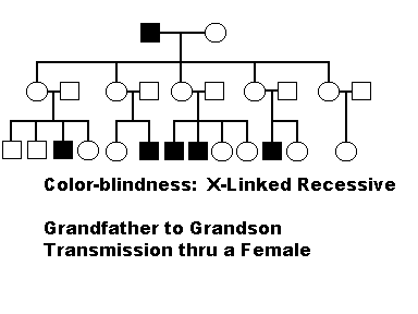
X- linked recessive

Characters-

  • Mostly males are sufferer of disease.
  • All male child developed from affected mother are diseased.
  • Female develop the disease only when her father is diseased and mother is carrier.
  • The trait is rare in pedigree.
  • The trait skips generations .


                             Image source-http://www231.pair.com/fzwester/courses/bis10v/week4/14xlinkeddisorders.html


Thank you for visiting my blog.Please feel free to share your comment on this article ,Please subscribe and share the articles to get more such articles


Tuesday, 24 May 2016

Apoptosis

Apoptosis- (Programmed cell death )It is a natural way in which  the cell kill itself. 

1.In situations like infection, inflammalation it is necessary.

2.In metastasis process of cancer to prevent the rapid growth of uncontrol cells apoptosis occur.
Changes -
1.Cell shrinkage 
2.Chromatin condensation
3.Membrane blebbing 
4.Nuclear collapse 

Types-

1.Intrinsic (the origin is from inner side of cell)
2.Extrinsic(pathway originated outside the cell)

1.  Intrinsic or mitochondrial pathway-

  • In a normal cell, the outer membranes of mitochondria display the protein Bcl-2 on their surface. Bcl-2 inhibits apoptosis.
  • Internal damage to the cell caused by protein Bax, to migrate to the surface of the mitochondria where it inhibits the protective effect of Bcl-2 and inserts itself into the outer mitochondrial membrane punching holes in it and causing cytochrome c to leak out.
  • The released cytochrome c binds to the protein Apaf-1 ("apoptotic protease activating factor-1") using the energy provided by ATP,these complexes aggregate to form apoptosomes.
  • The apoptosomes bind to and activate caspase-9.
  • Caspase-9 is one of a family of caspases which are all proteases. They get their name as they cleave proteins for example in aspartic acid (Asp) residues.
  • Caspase-9 cleaves and, in doing so, activates other caspases (caspase 3 and 7).
  • The activation of these  caspases creates an expanding cascade of proteolytic activity which leads to
    • digestion of structural proteins in the cytoplasm,
    • degradation of chromosomal DNA, and phagocytosis of the cell.

2.  The extrinsic or death receptor pathway-

  • Fas and the TNF receptor are integral membrane proteins with their receptor domains exposed at the surface of the cell 
  • Binding of complementary death activator (FasL and TNF respectively) transmits a signal to the cytoplasm that leads to-activation of caspase 8
  • Caspase 8 (like caspase 9) initiates a cascade of caspase activation leading to phagocytosis of the cell.
 When cytotoxic T cells recognize their target,
  • they produce more FasL at their surface.
  • This binds with the Fas on the surface of the target cell leading to its death by apoptosis.


                               Image source- http://www.nature.com/nrm/journal/v9/n1/fig_tab/nrm2308_F2.html
1.Apoptosis all are seen except
a necrosis
b. cell shrinking
c. nuclear clumping
d. inflammation                                                                          Ans.d


2.Starting point of apoptosis for programme cell death is -
a)Activation of endonuclease
b)Release of enzyme
c)Accumulation of calcium
d)Destruction by macrophages                                                    And.a


3.Chemotherapeutic drugs can cause:
A. Only necrosis
B. Only apoptosis
C. Both necrosis and apoptosis
D. Anoikis                                                                                 Ans.C


4.Annexin V assay is a method to detect?
a) Necrosis
b) Apoptosis
c) Inflammation
d) Neoplasia                                                                             Ans.B


5.Radiotherapy in tumor cells induces
a. Necrosis
b. Apoptosis
c. Phagocytosis 
d. Both necrosis and apoptosis                                                                      Ans.B(Radiation therapy causes apoptosis of tumors and surrounding tissue via free radical formation and dsDNA breakage.)


6.In apoptosis, Apaf-I is activated by release of which of
the following substances from the mitochondria?
(a) Bcl-2 
(b) Bax
(c) Bcl-XL
(d) Cytochrome C                                                                         Ans.D


7.Organelle that plays a pivotal role in apoptosis:
A. Endoplasmic reticulum
B. Golgi complex
C. Mitochondria
D. Nucleus                                                                                 Ans.C

8.Mechanism of mammalian apoptosis involves the most impor- tant role of the following protein:
A. Receptor for TNF 
B. BCL-2
C. TP53
D. CED-9                                                                                      Ans.B

NOTE-Apoptosis occurs in all ( Embryogenesis ,Menstruation ,Tumors ,Viral infection)

Thank you for visiting my blog.Please feel free to share your comment on this article ,Please subscribe and share the articles to get more such articles


Monday, 23 May 2016

ABC model

Homeiotic genes -It regulates the develpoment of anatomical structures in plants. The flowers of Arabidopsis (Brassicaceae family) consist of 4 concentric whorls .

Whorl 1=sepal
Whorl 2=petal
Whorl 3=stamen
Whorl 4=carpel

The expression of homeotic genes in 4 whorls results in production of transcription factors which control the phenotype of organs. Three classes of homeotic genes A,B and C determine organ identity by their expression in different whorls of cells.That is called ABC model.

In wild type flower (not mutant) -
 Whorl-1 (sepals) by the product of A genes (Apetala 1 and apetala 2)
 Whorl-2 (petals) by the product of A and B genes (apetala 3 and pistillata)
 Whorl-3 (stamens) by the product of B and C genes(agamous)
 Whorl 4 (carpels) by the action of C genes alone.

All except the Apetala 2 protein contain the same DNA- binding domain, the MADS box.
In mutant forms the role of these genes are deduced by inducing mutation in each of these genes.A and C genes are equally dominant .Mutation in A gene makes C gene more active. B gene always express in association with A and C genes.


ABC Model of flower development summary chart

(ABC model and changes associted with whorl formation due to mutation in A,B and C gene)

Image source-http://www.biologyexams4u.com/2015/03/abc-model-of-flower-development-in.html#.V0PPJpF97Dd


Q.Following are certain statements regarding the activities of homeiotic genes of classes A,B and C involved in floral oran identity-

a.Activity of A alone specifies sepals
b.Activity of B alone specifies petals
c.Activities of B and C form stamens
d.Activity of C alone specifies carpels
 Which one of the following combinations of above statements is correct?
1.A,B, and C                    3.A,B and D
2.B,C and D                     4.A,C and D

Ans. 4(A,C and D)

Thank you for visiting my blog.Please feel free to share your comment on this article ,Please subscribe and share the articles to get more such articles


Ramachandran plot

Ramachandran plot

It is also called as Ramachandran diagram discoverd by G.N.Ramachandran in 1963. It displays to visualize the distribution of conformational angle (dihedral angles ψ and φ) of amino acid residues in protein structure except glycine and proline.

The rotations of the polypeptide backbone around the bonds between N-Cα (called Phi, φ) and Cα-C (called Psi, ψ, see below for the graphics view of the angles). A special way for plotting protein torsion angles was also introduced by Ramachandran and co-authors, and was subsequently named the Ramachandran plot. The Ramachandran plot provides an easy way to view the distribution of torsion angles in a protein structure. It also provides an overview of excluded regions that show which rotations of the polypeptide are not allowed due to steric hindrance (collisions between atoms). The Ramachandran plot of a particular protein may also serve as an important indicator of the quality of its three-dimensional structures .

Torsion angles are among the most important local structural parameters that control protein folding - essentially, if we would have a way to predict the Ramachandran angles for a particular protein, we would be able to predict its fold. The torsion angles phi and psi provide the flexibility required for the polypeptide backbone to adopt a certain fold, since the third possible torsion angle within the protein backbone (called omega, ω) is essentially flat and fixed to 180 degrees. This is due to the partial double-bond character of the peptide bond, which restricts rotation around the C-N bond, placing two successive α-carbons and C, O, N and H between them in one plane. Thus, rotation of the protein chain can be described as rotation of the peptide bond planes relative to each other.


Since Gly is more flexible Ïˆ and Ï† combinations not tolerated. Proline, with the sidechain covalently linked to the preceding backbone N, is more tightly constrained than general  residues.  In the plot the φ values on the x-axis and the ψ values on the y-axis. The torsional angles determine the conformation of the residues and the peptide. Many of the angle  conformations are not possible because of steric hindrance.
 \mathrm{C^{\alpha}} - C is Ïˆ and  N-\mathrm{C^{\alpha}} is Ï†
Image source https://en.wikipedia.org/wiki/Ramachandran_plot
The  axis of the α-helix rotating in the y-plane. The Ramachandran plot of  peptide has points clustered about the values of φ= -57o and ψ= -47o which are the average values for α-helices. 
 Most β-sheets in globular proteins are twisted sheets which do not have flat parallel pleats. The Ramachandran plot of twisted sheet has points clustered about the values of φ= -130o and ψ= +140o which are the average values for twisted sheets.  


                     Image sourcehttps://www.studyblue.com/notes/note/n/protein-structure/deck/7778686
1.The area of allowed regions in the Ramachandran map will bw least for -
a.Gly                                c.L-ala
b.L-pro                             d.alpha-methylL-valine

Ans.L-pro 

2.JUNE 2017 CSIR 
The amino acids with Phi and psi values (-60,-40),(-59,-47) and (-80,120) will be adaopting which of the following confirmation ?
a.Helix-Helix-extended
b.Heix-coil-extended
c.Extended-extended-loop
d.Loop-loop-coil                                          Ans.A (Helix-Helix-extended)
Thank you for visiting my blog.Please feel free to share your comment on this article ,Please subscribe and share the articles to get more such articles

Friday, 20 May 2016

D- loop replication

D- loop replication-

The mitochondrial Dna  undergoes a process of replication called D- loop replication. It is found in circular Dna. Mitochondrial Dna is circular like plasmid. It is made up of 2 strands-

1.L-strand(light strand)
2.H-strand(heavy strand)

There are 2 different origin of replications ,one for light strand and one for heavy strand. The process of replication starts from origin of heavy strand. Replication in case of mitochondia is semiconservative . Heavy strand act as a leading strand and form displacememt loops .The loop expands as the process elongate. As a result of this a new light strand form by displacing the parental strand, whose replication structure is resembles to the letter D . When displaced strand passes the origin of replication of L-strand a new H -strand  synthesis starts. Because of replication structure D this mode of replication is called as D-loop replication of mitochondrial Dna.


Image source -http://slideplayer.com/slide/2758294/


Thank you for visiting my blog.Please feel free to share your comment on this article ,Please subscribe and share the articles to get more such articles.
Fluctuation test

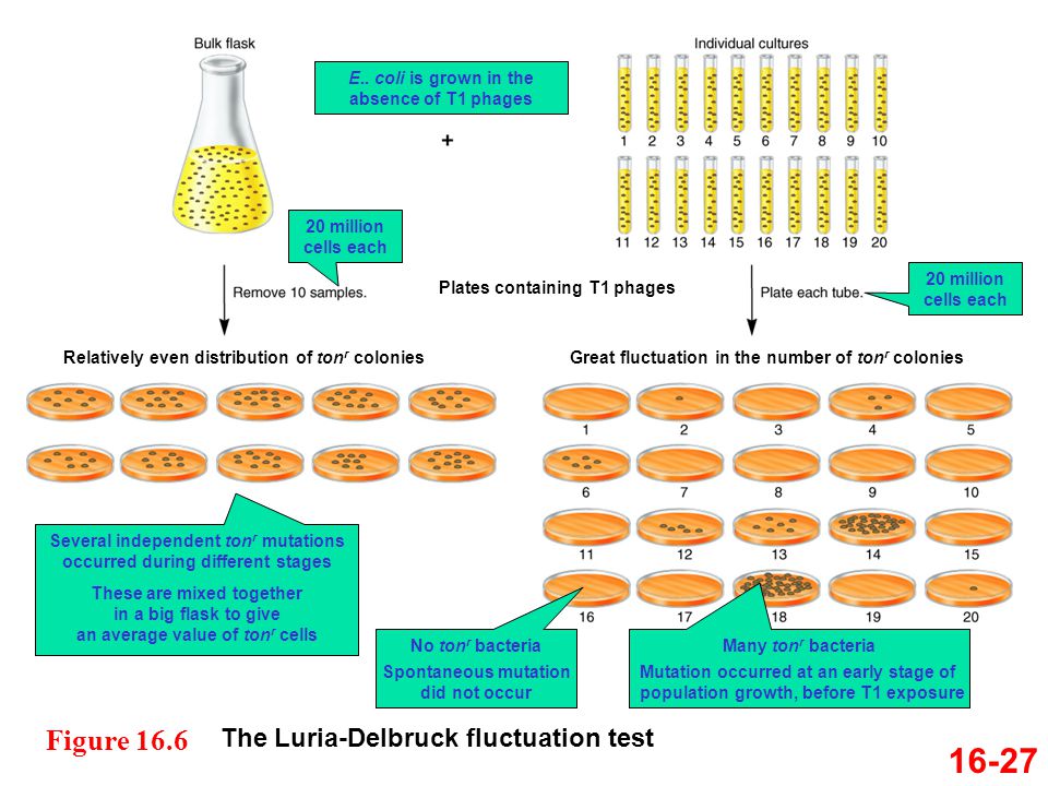
Fluctuation test- (by Luria and Delbruck) Distinguish between adaptive and spontaneous mutation.

  • It is a test to determine the randomness of mutation in a bacteria.
  • When bacteriophage T1 infects wild type E.Coli, it binds to a receptor named TonB in the outer membrane of the bacteria.
  •  Most of the bacteria are killed  by the insertion of phage  and new phages are released after replication .
  • When phage infects mutant type due to mutation in TonB gene altered receptor to which T1 can no longer bind and the cells are survive .
  •  Luria and Delbruck measured the number of mutants resistant to phage T1 in a large number of replicate cultures of E.coli. The numbers depend on how early during the growth period the first mutant cells arose.Early generation mutations give more mutant cells than late mutation.
  • The test is called fluctuation test as it measures the amount of fluctuation in the number of mutants found in replicate cultures.

                                            Image source -http://slideplayer.com/slide/2816451/


Thank you for visiting my blog.Please feel free to share your comment on this article ,Please subscribe and share the articles to get more such articles.

Thursday, 19 May 2016

Missence mutation

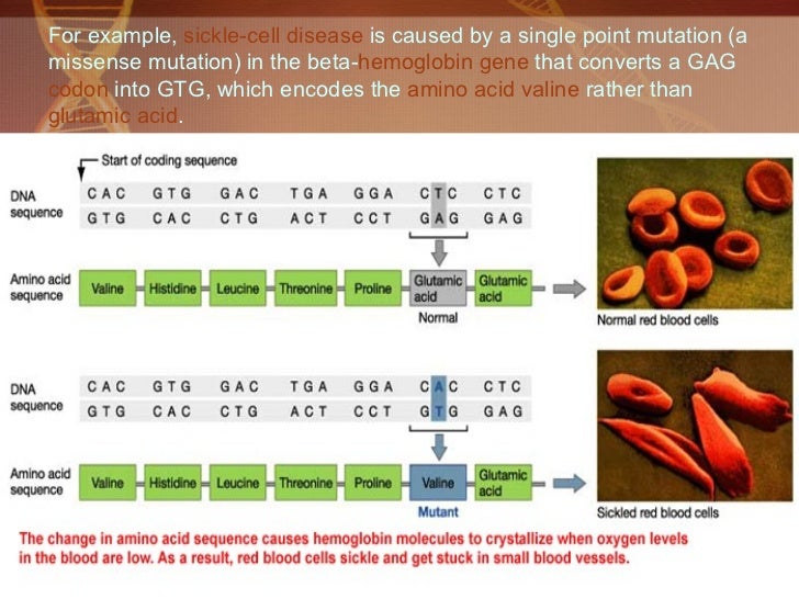
Missence mutation- Mutation  that alters the codon so that it specifies a different amino acid is know as missense mutation. The common example is sickle cell anemia.
  • In this the haemoglobin  which is oxygen transporting macromolecule present in the RBC of chordate animals undergoes missense mutation.
  • Haemoglobin A cosists of 2 identical alpha chains ( 141 amino acids) and 2 identical beta chains (146 amino acids).  As a result the  substituition of glutamic acid (GAG) to valine(GTG) takes place at aminoacid position 6 in the beta chain of haemoglobin .This changes the shape of the haemoglobin making it sickle shape due to change in shape of haemoglobin in oxygen unbound form.
For example,  sickle-cell disease  is caused by a single point mutation (a missense mutation) in the beta- hemoglobin   ge...
                                               Image source-http://www.slideshare.net/ShahabRiaz/genetic-disorders-2
MCQS.
1.The gene of sickle cell anaemia is inherited by
A) Blood cells
B) Bone cells
C) Sex chromosomes
D) Autosomes
ans. D

Thank you for visiting my blog.Please feel free to share your comment on this article ,Please subscribe and share the articles to get more such articles.


What is active site of an enzyme?

Active site-

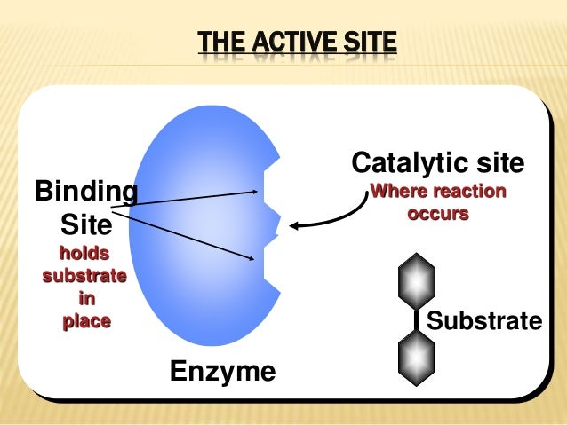
Definition- It is a binding site in enzyme  where the substrate binds to yield enzyme substrate complex and products by enhancing the chemical reaction.

The active site is usually a groove or pocket of the enzyme which can be located in a deep tunnel within the enzyme.

The active site fits with one specific type of substrate.Residues in the binding site form hydrogen bonds ,hydrophobic interactions, or temporary covalent interactions (vanderWaals) with the substrate to make an enzyme-substrate complex. 

Active site have 2 regions with unique  activities -

1.Binding site- Where the substrate binds.
2.Catalytic site- For catalytic chemical reaction.


                                    Image source-http://analytical.wikia.com/wiki/Active_site?file=Active_site.gif


Catalytic site
Where reaction
occurs
Binding
Site
holds
substrate
in
place Substrate
Enzyme
THE ACTIVE SITE

                              Image source-http://www.slideshare.net/AneettaDavis/simple-eutectic-systempb-ag


Thank you for visiting my blog.Please feel free to share your comment on this article ,Please subscribe and share the articles to get more such articles.

What are the properties of enzymes ?

Properties of enzymes -
                                                                                                                                                                                          
                                                                             Image source https://quizlet.com/21392287/biochemistry-chapter-11-flash-cards/                                                                                                                                                                 
  • Enzymes are bio-catalysts-Catalyst binds with reactants (called the substrate)accelerates the reactionand then repeatedly separates from the reaction products. 
  • Apoenzyme - The protein part of enzyme is called apoenzyme.
                               Apoenzyme+cofacter =Holoenzyme
  • Coenzyme- Coenzymes are non protein organic molecules used by enzymes to catalyse reactions. Many coenzymes cannot be synthesized in our body, they must be ingested as vitamins. NAD ,FAD and vitamin complexes are the examples of coenzyme.
  •  Cofacters-  Catalytically essential inorganic molecules or ions that are covalently bound to the enzyme,such as iron or magnesium. Participate in reaction by removing electrons ,protons and chemical groups from the substrate.
  • An enzyme  contain components that are not proteins, and these are grouped under prosthetic groups. Which includes (heme) present in hemoglobin and cytochrome.
  • All enzymes are proteins but all proteins are not enzymes.
  • Enzymes accelerate a reaction by decreasing energy required to form the transition state. 
1. Enzymes donot interfere with -
a. Free energy of reaction
b.Rate of reaction
c.Avtivation energy of transition state 
d. Reaction equilibrium 
Ans.   d  

Thank you for visiting my blog.Please feel free to share your comment on this article ,Please subscribe and share the articles to get more such articles.

Wednesday, 18 May 2016

What is frame shift mutation ?

 Frame shift mutation -
Types of mutations
Mutation
Point
mutation
Transition Transversions
Frame shift
Mutation
Insertions Deletions
Image source-http://www.slideshare.net/rajdangol303/mutation-35776184
  •  Mutation is a sudden change in nucleotide sequence.  
  •  It is the addition or deletion of base pair that occurs within the protein coding portion of a gene which  have the effect of shifting the translational reading frame.
  • In frame shift  mutation each codon acts as an open reading frame.
  • It  disrupts the translation changing the reading frame due to addition or deletion and Proteins are built incorrectly.

Frameshift Mutations             mRNA Normal                A U G    A A G U U U GGC GC A U UG C A A             Protein  ...

Image source -http://www.slideshare.net/openmichigan/081308d-ginsburgdnaseq-variation

Thank you for visiting my blog.Please feel free to share your comment on this article ,Please subscribe and share the articles to get more such articles. 


What is an ORF ?

Open Reading Frame-
  • It is a protein coding region consisting of the start codon and stop codon in DNA or RNA.
  • Stop codon is absent in open reading frame of prokaryotes as it haults the progress of the reading frame.
  • It comprises both the coding sequences exon and the non-coding sequences introns.
  • In enkaryotes , long ORFs can continue over non-translatable intron gaps, so spliced mRNA must be employed to determine ORFs. Short ORFs can occur outside genes – within the intron, segments of DNA outside genes that were formerly considered ‘junk’ DNA. 
  • A DNA open reading frame starts with ATG—coding for Met—in most species, and ends with a stop codon (TAA, TAG, or TGA).
Types of mutations in ORFs-

1.Nonsynonomous or mis-sense mutation 
2.Nonsense mutation
3.Synonomous or silent mutation 
4.Neutral non synonomous mutation 
5.Frameshift mutation (Deletion or insertion )


Thank you for visiting my blog.Please feel free to share your comment on this article ,Please subscribe and share the articles to get more such articles. 

Meselson and stahls experiment-

Meselson and Stahls experiment- 
(By density gradient centrifugation they did experiment to evaluate the 3 different models of replication.)
                          
  
   Hypothesis- Dna replicates semiconservatively.


  • E.coli bacteria cells were cultured (grown ) in a medium with 15N heavy isotope.  15N is a heavy isotope of nitrogen so the DNA synthesized after  Cscl density gradient centrifugation is of heavy density.
  • E. coli cells with only 15N in their DNA were transferred to a 14N medium . DNA was isolated  after successesive  replication cycles . 
  •  Replication cycle 1 (after 20 mins )- DNA was all of intermediate density in the 14N medium and  form a single band.This rules out the conservative replication model, which predicts that both heavy density DNA and light density DNA will be present, but no intermediate density will be present.
  •  Replication cycle  2- (after  40 mins ) - DNA with the semiconservative replication model as half the Dna are of intermediates (15N - 14N) and half  are of light ( 14N ) and this  eliminates the dispersive replication model.
  • After two replication cycles, two bands of DNA were seen, one of intermediate density and one of light density. 
  •  Result-  Semiconservative model predicts: half with 15N - 14N intermediate density DNA and half with 14N-14N light density DNA.( No intermediates)

Thank you for visiting my blog.Please feel free to share your comment on this article ,Please subscribe and share the articles to get more such articles.